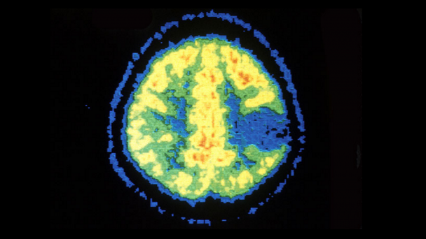
The FDA has approved an investigational new drug application for Tumor Paint BLZ-100, a protein-linked dye that highlights cancer cells in images so surgeons can precisely target brain tumors.The FDA move means Blaze Bioscience can proceed with clinical trials.
Twenty-one adult patients who need surgery for often-deadly glioma brain tumors are expected to enroll in the study, which is aimed at examining the safety of injecting the BLZ-100 molecule into the bloodstream, where it rushes to highlight cancer cells.
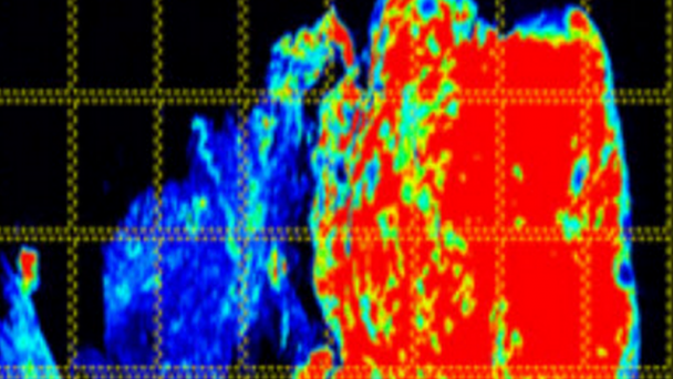
Images of a soft tissue sarcoma from a dog using Tumor Paint BLZ-100. Right: standard histological stain showing the morphology of the tissue; left: fluorescence provided by Tumor Paint, with cancerous cells highlighted in red. (Photo credit: Blaze Bioscience Inc.)
Press Release
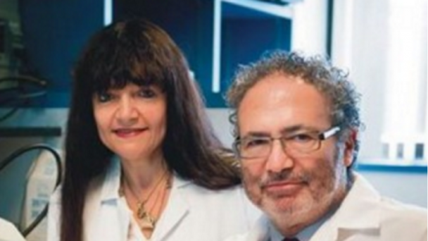
By JoNel Aleccia
Hutch News Sept. 26, 2014
A new protein-linked dye derived from scorpion venom that lights up cancer cells so surgeons can precisely target brain tumors will get a trial run in the U.S., Blaze Bioscience Inc. officials announced Thursday.
Food and Drug Administration officials have approved an investigational new drug application, or IND, for Tumor Paint BLZ-100, a molecule discovered and first developed by researchers at Fred Hutchinson Cancer Research Center, Seattle Children’s Hospital and the University of Washington.
“I think it really ...

