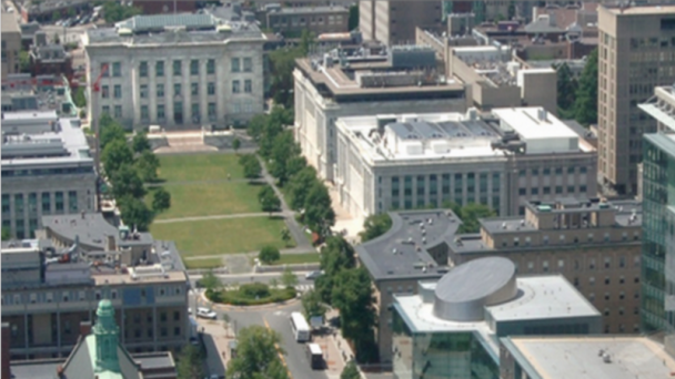
The Program in Neuroscience draws together neuroscientists from across Harvard. The physical home base of the program is located at the Longwood Campus of Harvard Medical School, in the Department of Neurobiology.
Research sites include the Longwood Medical Area, Cambridge Campus, Massachusetts General Hospital, and the McLean Hospital. The Center for Brain Science unites many neuroscience labs and houses in the newly established Swartz Program in Theoretical Neuroscience.
Web Information
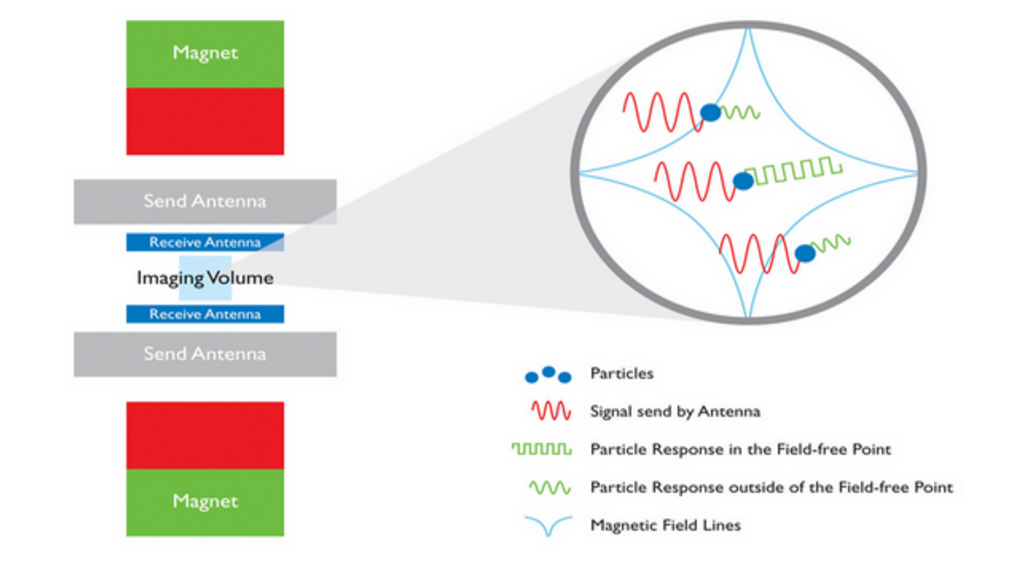
Website: dms.hms.harvard.edu/neuroscience/prospective/AboutPIN Research Sites: dms.hms.harvard.edu/neuroscience/prospective/ResearchSites Brain Initiative Grant – “Comprehensive Classification Of Neuronal Subtypes By Single Cell Transcriptomics” BRAIN Initiative Grant– “Magnetic Particle Imaging (MPI) for Functional Brain Imaging in Humans” BRAIN Initiative Grant– “Mapping neuronal chloride microdomains” Brain Initiative Grant– “Neural circuits in zebrafish: form, function and plasticity”
Contact Information
Email: karen_harmin@hms.harvard.edu Phone: (617) 432-0912 Address: Program in Neuroscience Harvard Medical School 220 Longwood Avenue Goldenson 129 Boston, MA 02115
About the Program in Neuroscience
Mission Statement: We are an inter-departmental Ph.D. program for training in neuroscience. Our mission is to provide students with the instruction, research experience, and mentoring they need to become leaders in research and education.
Who we are: The Program in Neuroscience draws together neuroscientists from across Harvard. The physical home base of the program is located at the Longwood Campus of Harvard Medical School, in the Department of Neurobiology. Most coursework occurs at this campus, ...
OnAir Post: Harvard Neuroscience


