Stanford Neurosciences Institute


Summary

The goal of the Stanford Neurosciences Institute is to understand how the brain gives rise to mental life and behavior.

The Institute’s interdisciplinary community of scholars will draw from a multiplicity of disciplines, including neuroscience, medicine, education, law and business.  Their discoveries aim to remodel understanding of brain function, individuals, and society, enabling positive change and enhancing human potential. Current research themes: The Changing Brain, Cracking the Neural Code, Enhancing the Brain, Understanding Thought, and How We Learn.

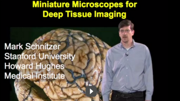
https://www.youtube.com/watch?v=v9s6W77jHHAVideo can’t be loaded because JavaScript is disabled: Cracking the Neural Code (https://www.youtube.com/watch?v=v9s6W77jHHA)

Information

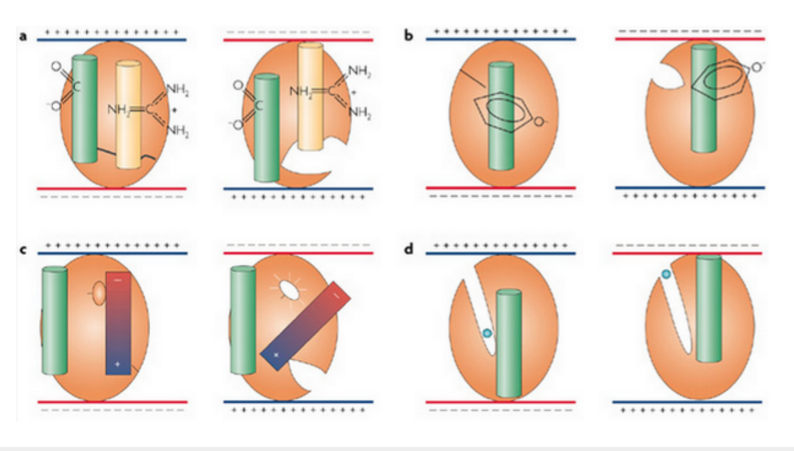
Website:  neuroscience.stanford.edu/ Brain Initiative Grant – “Protein voltage sensors: kilohertz imaging of neural dynamics in behaving animals”

Email: neuroscience@stanford.edu Phone: 650-497-8019 Address: James H. Clark Center 318 Campus Drive, Suite S170 Stanford, CA 94305-5443

Organization

Director: William T. Newsome


About Us

Our Mission

The goal of the Stanford Neurosciences Institute is to understand how the brain gives rise to mental life and behavior, both in health and in disease. Our research community draws from and informs multiple disciplines, including neuroscience, medicine, engineering, psychology, education and law. New discoveries will transform our understanding of the human brain, provide novel treatments for brain disorders, and promote brain health throughout the lifespan. We aim to create positive benefits ...

OnAir Post: Stanford Neurosciences Institute

William Newsome, PhD – Stanford


Director of the Stanford Neurosciences Institute and Professor of Neurobiology HHMI scientist Co-Chair, Advisory Committee to the NIH Director

Dr. Newsome’s research focuses on the neural mechanisms underlying visually based decision making and related issues in cognitive neuroscience. He seeks to understand how higher mammals acquire sensory information about the world, how that information is processed within the brain, and how behavioral responses to that information are organized.

Web Information

Stanford Webpage: med.stanford.edu/profiles/william-newsome

Lab Webpage: monkeybiz.stanford.edu/

HHMI Webpage: hhmi.org/scientists/william-t-newsome

Contact Information

Email: bnewsome@stanford.edu

Phone: (650) 725-5814

Address: Stanford School of Medicine 291 Campus Drive Li Ka Shing Building Stanford, CA 94305-5101

Research Interests

From lab page

The long-term goal of our research is to understand the neuronal processes that mediate visual perception and visually guided behavior. To this end we are conducting parallel behavioral and physiological experiments in animals that are trained to perform selected perceptual or eye movement tasks. By recording the activity of cortical neurons during performance of such tasks, we gain initial insights into the relationship of neuronal activity to the animal’s behavioral capacities. Hypotheses concerning this relationship are tested by modifying neural activity within local cortical circuits to determine whether behavior is effected in a predictable manner. Computer modelling techniques are then used to develop more refined hypotheses concerning the relationship of ...

OnAir Post: William Newsome, PhD – Stanford

Karl Deisseroth, MD, PhD – Stanford

D.H. Chen Professor of Bioengineering and of Psychiatry and Behavioral Sciences,Stanford University Howard Hughes Medical Institute Investigator Member of the Advisory Committee to the NIH Director

Deisseroth focuses on developing molecular and cellular tools to observe, perturb, and re-engineer brain circuits. His lab employs a range of techniques including neural stem cell and tissue engineering methods, electrophysiology, molecular biology, neural activity imaging, animal behavior, and computational neural network modeling. As a clinician in the psychiatry department, Dr. Deisseroth employs novel electromagnetic brain stimulation techniques.

Web Information

School of Medicine Webpage: med.stanford.edu/profiles/karl-deisseroth

Lab Webpage: stanford.edu/group/dlab/about_pi

HHMI Webpage: hhmi.org/scientists/karl-deisseroth

Wikipedia Entry: wikipedia.org/wiki/Karl_Deisseroth

Contact Information

Email:deissero@stanford.edu

Address: 318 Campus Drive West Clark Center W083 Department of Bioengineering Stanford University Stanford, CA 94305

Research Summary

From HHMI page

Karl Deisseroth develops optical methods for high-resolution investigation of intact biological systems. His group has pioneered optogenetics, a technology that uses light to control millisecond-precision activity patterns in defined cell types in the brains of freely moving mammals, and CLARITY, a chemical engineering technology that enables high-resolution structural and molecular access to intact brains. A practicing psychiatrist, Deisseroth has also applied his technologies to study anxiety, depression, and social dysfunction.

Our research group builds optical tools for precise, high-resolution investigation of intact biological systems, with a focus on the vertebrate central nervous system; in particular, ...

OnAir Post: Karl Deisseroth, MD, PhD – Stanford

Mark J Schnitzer, PhD – Stanford


Associate Professor of Biology and Applied Physics, Stanford HHMI Investigator Principal Investigator, Schnizer Group

Dr. Schnitzer has longstanding interests in neural circuit dynamics and optical imaging focusing on: the development and application of fiber-optic, micro-optic, and nanophotonic imaging techniques for studies of learning and memory; in vivo fluorescence imaging and behavioral studies of hippocampal-dependent cognition and learning; and development of high-throughput, massively parallel imaging techniques for studying brain function in Drosophila.


Web Information

Webpage: stanford.edu/dept/app-physics/cgi-bin/person/schnitzer-mark-j/ Stanford School of Medicine webpage: med.stanford.edu/profiles/mark-schnitzer Stanford Neurosciencs Institute Brain Initiative Grant

Contact Information

Email: mschnitz@stanford.edu Phone: 650) 723-4027 Address: James H. Clark Center – Room W080 318 Campus Drive Stanford, CA 94305


Biography

Education

Harvard University Cambridge, MA A.B. summa cum laude 1988-1992 Physics

Cambridge University Cambridge, UK Certificate 1992-1993 Mathematics Princeton University Princeton, NJ M.A. 1993-1994 Physics

Princeton University Princeton, NJ Ph.D. 1994-1999 Physics (advisor: Prof. Steven M. Block)

Positions and Honors

2008-present Investigator, Howard Hughes Medical Institute; Stanford University.

2006-present Janelia Farm Research Campus, Howard Hughes Medical Institute, Scientific Visitor Program, Ashburn VA.

2003-present Assistant Professor, Dept. of Applied Physics and Dept. of Biological Sciences; Faculty Member, Neuroscience Program, Biophysics Program, Stanford Univ., Stanford, CA.

1999-2003 Member of Technical Staff, Physical Sciences Laboratory, Biological Computation Research Department, Bell Laboratories, Lucent Technologies, Murray Hill, NJ.

1994-1999 Ph.D. Research, with Steven M. Block, Dept. of Molecular Biology, ...

OnAir Post: Mark J Schnitzer, PhD – Stanford

Henry T. Greely, JD – Stanford

Professor of Law, Stanford University Director, Center for Law and the Biosciences Director, Stanford Program in Neuroscience and Society (SPINS) At large member, BRAIN Initiative Multi-Council Working Group

Henry Greely specializes in the ethical, legal, and social implications of new biomedical technologies, particularly those related to neuroscience, genetics, or stem cell research.

Web Information

Stanford Webpage:  www.law.stanford.edu/profile/henry-t-greely

SPINS website: https://law.stanford.edu/stanford-program-neuroscience-society/

Stanford Center for Law and the Biosciences (CLB) website: https://law.stanford.edu/center-for-law-and-the-biosciences/

International Neuroethics Society founders: http://www.neuroethicssociety.org/who-are-we

Blog:  blogs.law.stanford.edu/lawandbiosciences/

Twitter: @HankGreelyLSJU

Contact Information

Email: hgreely@stanford.edu

Phone:  650 723.2517

Address: Neukom Building Room N361

BRAIN Blog posts

Turn right at the cerebellum: President Obama maps the brain

Feb. 21, 2013 by Amanda Rubin

This week, the New York Times reported on a new Obama initiative that, in comparison to gun control or the economy, might seem a little frivolous. It’s called the “Brain Activity Map.”

Three Billion Dollars

The name of the project says it all: The goal is to map the connections in the brain in the same way the Human Genome Project mapped out the genes in human DNA. It’s expected to cost about $3 billion dollars over ten years.

If that seems like a pretty heavy price tag for the American people to take on, especially now, just to let scientist go ...

OnAir Post: Henry T. Greely, JD – Stanford

Miniature Microscopes for Deep Tissue Imaging

https://www.youtube.com/watch?v=C1HO3ot0K00Video can’t be loaded because JavaScript is disabled: Microscopy: Miniature Microscopes for Deep Tissue Imaging (Mark Schnitzer) (https://www.youtube.com/watch?v=C1HO3ot0K00)

This lecture describes recent work on developing small microscopes for deep tissue imaging that can surgically implementing into living and awake animals. Exciting applications are described for imaging the activity and long term shape changes of single neurons in the brain.

Video published on Nov. 11, 2013 by iBioEducation

Mark Schnitzer Profile

Associate Professor of Biology and Applied Physics, Stanford HHMI Investigator Principal Investigator, Schnizer Group

Dr. Schnitzer has longstanding interests in neural circuit dynamics and optical imaging focusing on: the development and application of fiber-optic, micro-optic, and nanophotonic imaging techniques for studies of learning and memory; in vivo fluorescence imaging and behavioral studies of hippocampal-dependent cognition and learning; and development of high-throughput, massively parallel imaging techniques for studying brain function in Drosophila.

OnAir Post: Miniature Microscopes for Deep Tissue Imaging

OpenfMRI enables sharing brain research data

https://www.youtube.com/watch?v=7HBusVeG8AQ

“OpenfMRI allows neuroscientists to share brain research data”

Researchers around the world can compare notes on one of the most powerful tools available for imaging human brain function, the fMRI, thanks to support from the National Science Foundation (NSF). An fMRI is a functional magnetic resonance imaging scan that measures brain activity by detecting changes in blood oxygenation and flow.

NSF BRAIN Initiative Science Nation – March 15, 2015

OnAir Post: OpenfMRI enables sharing brain research data

Schnitzer Group – Stanford

Principal Investigator: Mark J Schnitzer Stanford Neurosciencs Institute

The Schnitzer Group has three major research efforts: Development and application of fiber-optic, micro-optic, and nanophotonic imaging techniques for studies of learning and memory in behaving mice and for clinical uses in humans; In vivo fluorescence imaging and behavioral studies of hippocampal-dependent cognition and learning; and Development of high-throughput, massively parallel imaging techniques for studying brain function in large numbers of Drosophila concurrently.


Web Information

Website:  http://pyramidal.stanford.edu/index.html Brain Initiative Grant

Contact Information

Email: schnizerlab@gmail.com Address: James H. Clark Center – Room W080 318 Campus Drive Stanford, CA 94305

Research

Dr. Schnitzer has longstanding interests in neural circuit dynamics and optical imaging, and his laboratory has three major research efforts:

In vivo fluorescence imaging and behavioral studies of cerebellar-dependent motor control and motor learning. Development and application of fiber-optic fluorescence microendoscopy imaging techniques for studies of learning and memory in behaving mice and for clinical uses in humans. Development of high-throughput, massively parallel imaging techniques for studying brain function in large numbers of Drosophila concurrently.

The long-term goal of our research is to advance experimental paradigms for understanding normal cognitive and disease processes at the level of neural circuits, with emphasis on learning and memory processes. By contrast, much current research on learning and memory concentrates on ...

OnAir Post: Schnitzer Group – Stanford

Protein voltage sensor imaging in vivo

Principal Investigator: Mark J Schnitzer Stanford Neuroscience Title: “Protein voltage sensors: kilohertz imaging of neural dynamics in behaving animals” BRAIN Category: Large-Scale Recording-Modulation – Optimization (RFA NS-14-008)

Dr. Schnitzer and his team have created a new system for developing optical voltage sensors, which will allow scientists to simultaneously record firing of large groups of neurons or electrical activity in precise locations inside of neurons, such as synapses.

NIH Webpages

The diagrams show a hypothetical protein (orange oval) and the formation of an active site, which is due to a voltage-induced conformational change that is mediated by the defined regions of the protein (green and yellow cylinders). a | Charged amino acids may move within membranes in response to changes in voltage. The side groups of Asp and Arg are shown. b | Reorientation of an intrinsic residue dipole, such as Tyr, through changes in the field. c | An alpha-helix that is the length of the membrane (red to blue gradient) has a dipole moment that is equivalent to the length of the helix that separates plusminus0.5 electronic charges (e0); therefore, it can also reorientate when the field is changed. The oval that is attached to the alpha-helix ...

OnAir Post: Protein voltage sensor imaging in vivo

Skip to toolbar