Magnetic Particle Imaging (MPI)

Principal Investigator: Lawrence Wald Neuroscience@Harvard Title: “Magnetic Particle Imaging (MPI) for Functional Brain Imaging in Humans” BRAIN Category: Next Generation Human Imaging (RFA MH-14-217)

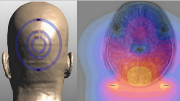
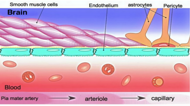
The Wald team plans to use an iron-oxide contrast agent to track blood volume, which will permit dramatically more sensitive imaging of human brain activity than existing methods.

NIH Webpages

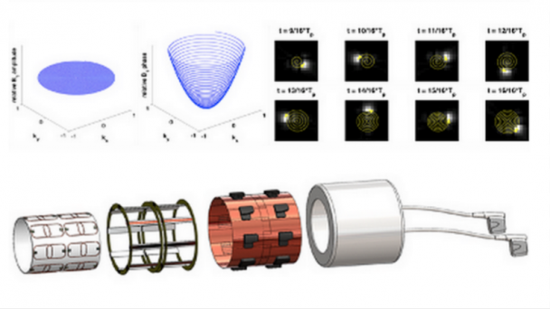
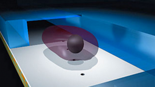
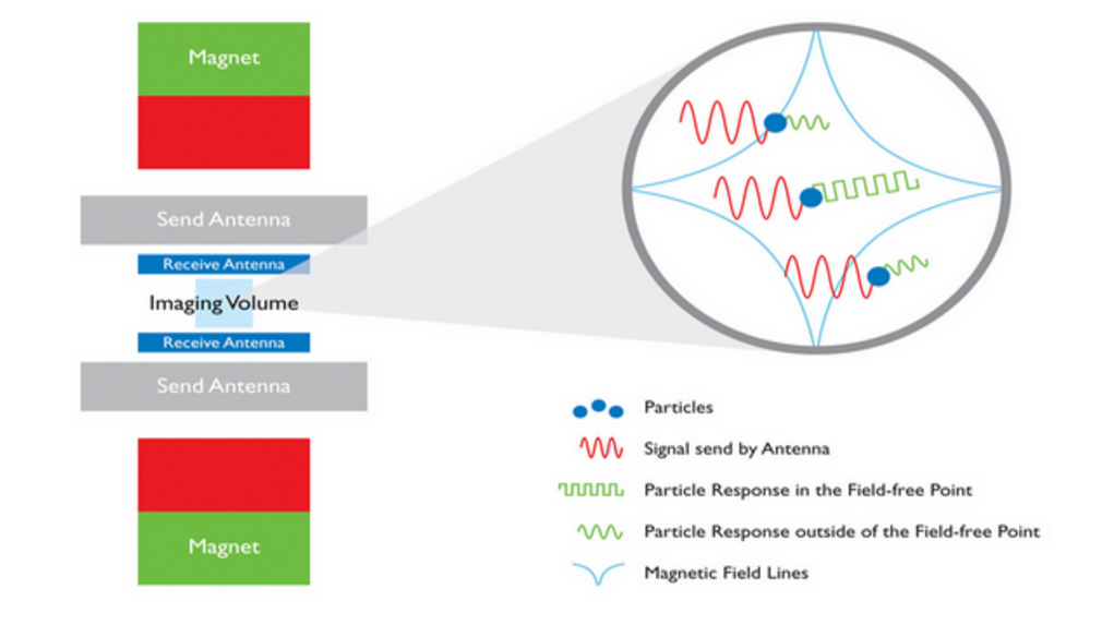
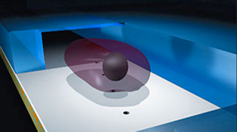
Schematic set up and operating principle of the Magnetic Particle Imaging technology. Phillips MPI.

Project Description

In this planning grant we propose several engineering developments to advance Magnetic Particle Imaging (MPI) to replace MRI as the next-generation functional brain imaging tool for human neuroscience. We assemble a group of technology experts to solve a myriad of identified and unidentified barriers, we employ simulation and bench-top experiments to characterize and test solutions for these technical obstacles and validate solutions by bench testing specific sub-sections of the imager. Finally we simulate the overall performance of the planned device and assess its benefit for human functional brain imaging. MPI is a young but extremely promising technology that uses the nonlinear magnetic response of iron- oxide nanoparticles to localize their presence in the body. MPI directly detects the nanoparticle’s magnetization rather than using secondary effects on the Magnetic Resonance relaxation times. ...

OnAir Post: Magnetic Particle Imaging (MPI)

Imaging Brain Function with Portable MRI

Principal Investigator: Michael Garwood Institute for Translational Neuroscience, University of Minnesta Title: “Imaging Brain Function in Real World Environments & Populations with Portable MRI” BRAIN Category: Next Generation Human Imaging (RFA MH-14-217)

By employing smaller, less cumbersome magnets than used in existing MRI, Dr. Garwood and colleagues will create a downsized, portable, less expensive brain scanner.

NIH Webpages

Project Description

Functional magnetic resonance imaging (fMRI) continues to play a critical role in understanding the human brain. Yet current fMRI technology is far less than ideal for studying brain function due to the unnatural environment and restricting space of the magnet bore. Furthermore, fMRI cannot be performed on subjects who have metallic implants in their body (e.g., the elderly, soldiers and veterans), or who are impaired by certain physical disabilities as occurs in a variety of neurological and vestibular disorders. Finally, due to its expense and infrastructure requirements, MRI’s predominant accessibility to wealthier institutions has resulted in a highly biased subject sampling and a shortage of studies in non-western environments and cultures. The general methodology used to obtain MR images today is essentially the same as that used approximately 4 decades ago. One major drawback of such methodology is that the tolerated magnetic field variation over the brain is ...

OnAir Post: Imaging Brain Function with Portable MRI

Advancing MRI & MRS Technologies

Principal Investigator: Wei Chen Institute for Translational Neuroscience, University of Minnesota Title: “Advancing MRI & MRS Technologies for Studying Human Brain Function and Energetics” BRAIN Category: Next Generation Human Imaging (RFA MH-14-217)

Dr. Chen’s team will achieve unprecedented higher resolution magnetic resonance imaging and spectroscopy scanning by integrating ultra-high dielectric constant material and ultra-high-field techniques.

NIH Webpages

Engineering for Ultrahigh field Magnetic Resonance Imaging (MRI) and Spectroscopy (MRS)

Project Description

Magnetic resonance (MR) imaging (MRI) and in vivo MR spectroscopy (MRS) techniques have become indispensable tools for imaging brain structure, function, connectivity, neurochemistry and neuroenergetics, and for investigating neurological disorders. However, it remains a challenge to achieve superior MRI/MRS detection sensitivity, spatial and temporal imaging resolutions adequate for addressing fundamental and challenging neuroscience questions even with the most advanced technology. The prevailing paradigms for improving MRI/MRS performance largely invoke increasing the magnetic field strength, which may have reached practically achievable limits for human studies due to many technological and safety (i.e., high specific absorption rate (SAR)) concerns, and increasing the receiver channel count which is also ultimately limited due to noise characteristics of coils of decreasing size. To alleviate these major limitations, this R24 proposal relies on the interdisciplinary research efforts and ...

OnAir Post: Advancing MRI & MRS Technologies

MRI Neuro-Electro-Magnetic Oscillations

Principal Investigator: Allen W Song Duke Institute for Brain Sciences Title: “Path Toward MRI with Direct Sensitivity to Neuro-Electro-Magnetic Oscillations” BRAIN Category: Next Generation Human Imaging (RFA MH-14-217)

Dr. Song’s group will develop a scanner technology sensitive enough to image brain activity in high resolution by directly tuning in the electromagnetic signals broadcast by neurons.

NIH Webpages

Spiral imaging is a fast MRI technique that is widely used in functional MRI. It is, however, vulnerable to spatial and temporal variations of the static magnetic field (B0) caused by susceptibility effects, subject motion, physiological noise, and system instabilities, resulting in blurring artifacts. To address these issues, we have developed a novel off-resonance correction method, based on k-space energy spectrum analysis (KESA), for inherent and dynamic B0 mapping and deblurring in spiral imaging. This method can generate a B0 map from the k-space data at each time point, without requiring any additional data acquisition or pulse sequence modification, and correct for the blurring caused by both spatial and temporal B0 variations, resulting in a high spatial and temporal fidelity.

Project Description

In response to the NIH RFA-MH-14-217 on “Planning for Next Generation Human Brain Imaging”, we propose a comprehensive plan to organize the ...

OnAir Post: MRI Neuro-Electro-Magnetic Oscillations

Vascular Interfaces for Brain Imaging

PI: Robert Desimone Massachusetts Institute of Technology Title: “Vascular Interfaces for Brain Imaging and Stimulation” BRAIN category: Next Generation Human Imaging (RFA MH-14-217)

Dr. Desimone’s project will access the brain through its network of blood vessels to less invasively image, stimulate and monitor electrical and molecular activity than existing methods.

NIH Webpages

Sketch showing constitution of blood vessels inside the brain. Credit: Armin Kübelbeck

Project Description

Functional MRI (fMRI), EEG, and other completely noninvasive modalities for large-scale imaging of human brain activity have pioneeringly revealed many human brain functions, but cannot reach the single-neuron, single-spike level of neural code analysis possible in animals obtained using electrodes. This is partly due to the indirect methods of observation employed (e.g., blood flow for fMRI) and due to blurring of signals over distance by the skull (e.g., for EEG). In contrast, invasive approaches such as trans-cranially implanted multi- electrode arrays can achieve single-cell, single-spike resolution, but they necessitate opening of the skull – and, for implanted arrays, damage of the brain tissue – limiting utility to a small fraction of the population, those undergoing neurosurgery for some intractable brain disorder that justifies the risk. Trans-cranially implanted arrays also degrade i performance over time ...

OnAir Post: Vascular Interfaces for Brain Imaging

Ultrasonic neuromodulation in vivo

PI: Doris Ying  Tsao California Institute of Technology Title: “Dissecting human brain circuits in vivo using ultrasonic neuromodulation” BRAIN category: Next Generation Human Imaging (RFA MH-14-217)

In rodents, monkeys and eventually humans, Dr. Tsao’s team will explore use of non-invasive, high resolution ultrasound to impact neural activity deep in the brain and modify behavior.

NIH Webpages


Project Description

A dream of neuroscience is to be able to non-invasively modulate any given region of the human brain with high spatial resolution. This would open new horizons for understanding human brain function and connectivity, and create completely new options for the non-invasive treatment of brain diseases such as intractable epilepsy, depression, and Parkinson’s disease. Current non-invasive brain stimulation methods such as transcranial magnetic stimulation (TMS) and transcranial electrical stimulation (TES) can be applied only to superficial cortical areas, with crude 1 cm-scale resolution, limits placed upon these techniques by fundamental physics. Ultrasonic neuromodulation, the use of ultrasound as an energy modality to affect the activity of the brain, could overcome these limitations and thereby transform both basic and clinical human neuroscience. In fact, the engineering challenge of non-invasively focusing ultrasound to mm-sized regions, either shallow or deep in the brain, has been solved: clinical studies have already demonstrated ...

OnAir Post: Ultrasonic neuromodulation in vivo

Micro-Dose, Wearable PET Brain Imager

Principal Investigator:  Julie Brefczynski-Lewis WVU Center for Neuroscience Title: Imaging the Brain in Motion: The Ambulatory Micro-Dose, Wearable PET Brain Imager BRAIN Category: Next Generation Human Imaging (RFA MH-14-217)

Dr. Brefczynski-Lewis and co-workers will engineer a wearable PET scanner that images activity of the human brain in motion – for example, while taking a walk in the park.

NIH Webpages

Brefczynski-Lewis, assisted by graduate student Chris Bauer, dons her PET-helmet prototype to demonstrate the device’s portability. (Lois Raimondo/For The Washington Post)

Project Description

Our vision is to design the first truly mobile molecular brain imager that can be used on healthy subjects to study the functioning of the human brain during motion. The ultimate goal is to be able to image subjects during a proverbial “walk in the park” and other natural activities. We selected PET technology as the most likely to succeed in the next decade to provide the desired functionality of such a brain imager. While MRI is an exceptionally powerful and versatile imaging modality, and there are even upright MRIs for structural brain scans, for functional fMRI scans the subjects must stay still and in horizontal position inside a narrow bore of a strong-field MRI magnet. What we ...

OnAir Post: Micro-Dose, Wearable PET Brain Imager

Multiplexed Nanoscale In Situ Proteomics

Edwards S. Boyden Massachusetts Institute of Technology Title: “Ultra-Multiplexed Nanoscale In Situ Proteomics for Understanding Synapse Types” BRAIN category: Tools for Cells and Circuits (RFA MH-14-216)

Dr. Boyden’s team will simultaneously image both the identities and locations of multiple proteins within individual synapses – made possible by a new technique called DNA-PAINT.

NIH Webpages

DNA-PAINT super-resolution image of microtubules inside a fixed HeLa cell using Atto 655–labeled imager strands (10,000 frames, 10-Hz frame rate). Inset, labeling and imaging schematic for DNA-PAINT in a cellular environment. From Neuron doi:10.1038/nmeth.2835

Project Description

Significant work is ongoing to reveal how different cell types in the brain contribute to behavior and pathology, and how they change in plasticity and disease, empowered by new genetic, optical, and physiological tools. However, the functional activity and dysregulation of neuronal circuits relies critically on the in situ molecular composition of neuronal synapses. Although it is clear that the properties of a given synapse are determined by, amongst other things, the specific types of cells that are thus connected, far less is known about the diversity of synapse types in the brain than cell types, perhaps because this is an intrinsically proteomic problem: a given neuron might make many ...

OnAir Post: Multiplexed Nanoscale In Situ Proteomics

Enhancers define cortical interneuron types

Principal Investigator: John L. R. Rubenstein UCSF Neuroscience Title: “Identification of enhancers whose activity defines cortical interneuron types” BRAIN Category: Tools for Cells and Circuits (RFA MH-14-216)

Dr. Rubenstein and colleagues plan to identify enhancer molecules specific to particular types of interneurons – that relay neural signals – and use this information to profile distinct cell types and new ways to manipulate genes.

NIH Webpages

Cortical Circuits: Projection Neurons and Interneurons

Project Description

Molecular definitions of neural cell types largely depend on the expression of RNAs or proteins as assessed by in situ hybridization, RNA array and sequencing, and immunohistochemistry. However, recent studies are demonstrating that gene regulatory elements, such as enhancers, can have highly specific spatial and temporal activity patterns in the developing brain. Thus, enhancer activity can be used to define neural cell types, and importantly, also have other broad applications. First, they can be used as tools to drive gene expression in specific cell types, which can then be used to visualize and/or purify the cells (GFP), modify gene expression in the cells (Cre), modify electrical activity (channel rhodopsin), and visualize electrical activity in the cells (GCaMP). Secondly, knowledge about the nature and position of enhancers enables geneticists ...

OnAir Post: Enhancers define cortical interneuron types

Remote regulation of neural activity

The Stanley team will focus on the development of tools to instantly and precisely target cell activity deep in the brain using radio waves, nanoparticles and genetically modified viruses.

NIH Webpages

OnAir Post: Remote regulation of neural activity

Holographic optogenetics and olfactory coding

Principal Investigator: Dmitry Rinberg NYU Neuroscience Institute Title: “Behavioral readout of spatiotemporal codes dissected by holographic optogenetics” BRAIN Category: Understanding Neural Circuits (RFA NS-14-009)

Dr. Rinberg’s team aims to understand how the brain turns odors into nerve signals by activating and recording neurons in the olfactory bulbs of mice as they detect a variety of odors.

NIH Webpages

Scientists are now able to determine which receptors respond to certain odors in awake, freely behaving animals. This image is for illustrative purposes only and shows a coronal section through the main olfactory bulb of an adult male mouse. Credit Matt Valley.

Project Description

Two of the most fundamental questions of sensory neuroscience are: 1) how is stimulus information represented by the activity of neurons at different levels of information processing? And 2) what features of this activity are read by the higher brain areas to guide behavior? The first question has been the subject of a large body of work across different sensory modalities. To answer the second question, one needs to establish a causal link between neuronal activity and behavior. In many systems, fine spatiotemporal patterns of activity underlie the neural representation of information. In these systems, deciphering the salient ...

OnAir Post: Holographic optogenetics and olfactory coding

Nontoxic transsynaptic tracing

Principal Investigator: Ian Wickersham MIT Neuroscience Title: “Novel technologies for nontoxic transsynaptic tracing” BRAIN Category: Tools for Cells and Circuits (RFA MH-14-216)

Dr. Wickersham and colleagues will develop nontoxic viral tracers to assist in the study of neural circuitry underlying complex behaviors.

NIH Webpages

(a–c) Injection site in mouse somatosensory thalamus of an equal mixture of two deletion-mutant rabies viral vectors: VSVG-enveloped vector encoding mOrange2 (yellow), and an RVG-enveloped vector encoding mCherry (red). The VSVG-enveloped vector prolifically infects thalamic neurons at the injection site, whereas the RVG-enveloped one infects far fewer cells locally. (d–f) In the somatosensory cortex of the same mouse, mOrange2-filled thalamocortical axons ascend to layer 4 and densely ramify among the apical dendrites of mCherry-filled layer 6 corticothalamic cells retrogradely infected by the co-injected retrograde virus. Scale bar, 200 μm, applies to all panels.

Project Description

Genetic tools have dramatically increased the power and resolution of neuroscientific experiments, allowing monitoring and perturbation of specific neuronal populations within the brain, often in the context of complex cognitive and behavioral paradigms. However, the usefulness of these tools is limited by the available means of delivering them in circuit-specific ways, a major drawback in view of the critical importance of specific connectivity ...

OnAir Post: Nontoxic transsynaptic tracing

Optogenetic toolkit for control of cells

PI: Gregory Hannon,  Hannon Lab Institution: Cold Spring Harbor Laboratory Title: “An optogenetic toolkit for the interrogation and control of single cells.” BRAIN Category: Tools for Cells and Circuits (RFA MH-14-216)

Dr. Hannon’s group will develop optogenetic techniques that use pulses of light to control genes and isolate proteins in specific cell types in the brain for molecular studies.

Project Description

Our understanding of brain function at the cellular and circuit level is critically dependent on the ability to interrogate and alter neural cells withhigh specificity. The use of light, either through single-photon or multi- photon excitation, is the onl method that provides sufficient resolution to probe the brain at the cellular and subcellular levels. While light-activated molecules, like optogenetic proteins or photocaged compounds, have allowed many key insights in neuroscience, their use is still limited to those processes that can be affected by membrane channels. We propose to develop a toolkit allowing the interrogation, regulation, and modification of genetic information in brain cells using light. We wll build on a technology we have recently developed, “LaserTag”, based on the light-dependent interaction between protein tags (i.e. SNAP-tag or HALO-tag) and caged chemical ligands. Such interaction can be either use to recover molecules through affinity purification or to force the ...

OnAir Post: Optogenetic toolkit for control of cells

Activity measurement at single cell

Principal Investigator: Craig Forest Georgia Institute of Technology Title: “In-vivo circuit activity measurement at single cell, sub-threshold resolution” BRAIN Category: Tools for Cells and Circuits (RFA MH-14-216)

Dr. Forest’s team will use a newly developed robot guided technique to measure precise changes in electrical activity from individual neurons that are connected over long distances across the brain, to understand how these connections change when our brains go into different states, such as sleeping and waking.

NIH Webpages


Whole-cell patch clamp electrophysiology of neurons, although a gold standard technique for high-fidelity analysis of the biophysical mechanisms of neural computation and pathology, requires great skill to perform. We have developed a simple robot that automatically performs patch clamping in vivo, algorithmically detecting cells by analyzing the temporal sequence of electrode impedance changes. We demonstrate good yield, throughput, and quality of recording in mouse cortex and hippocampus..

Project Description

Neurons communicate information through fluctuations in the electrical potentials across their cellular membranes. Whole-cell patch clamping, the gold standard technique for measuring these fluctuations, is something of an art form, requiring great skill to perform on only a few cells per day. Thus, it has been primarily limited to in vitro experiments, a few in ...

OnAir Post: Activity measurement at single cell

Dreadd2.0: A Chemogenetic Toolkit

Principal Investigator: Bryan L Roth UNC Neuroscience Title: ” Dreadd2.0: An Enhanced Chemogenetic Toolkit” BRAIN Category: Tools for Cells and Circuits (RFA MH-14-216)

Dr. Roth and colleagues will build second generation technology that uses artificial neurotransmitters and receptors to manipulate brain activity simultaneously across select cells and pathways to understand their functions and potentially treat brain disorders.

NIH Webpages

Roth’s Lab has pioneered the use of directed molecular evolution to create GPCRs which are suitable for remotely controlling cellular signaling. Using a variety of mouse genetic approaches (e.g. Cre-mediated recombination. they are able to control neuronal firing and non-neuronal signaling in real-time in awake, freely moving animals.Ongoing projects are to use this technology to deconstruct the neuronal requirements for simple and complex behaviors, particularly as they relate to schizophrenia and drug abuse

Project Description

The Brain Research through Advancing Innovative Neurotechnologies (BRAIN) Initiative has the ambitious goal of elucidating how neuronal ensembles interactively encode higher brain processes. To accomplish this goal, new and improved methods for both recording and manipulating neuronal activity will be needed. In this application, we focus on technologies for manipulating neuronal activity. The major significance of this application is that we will provide an enhanced chemogenetic toolbox that ...

OnAir Post: Dreadd2.0: A Chemogenetic Toolkit

Mapping Sensory-Motor Pathways

Principal Investigator: Michael Dickinson Caltech Neuroscience Title: “Integrative Functional Mapping of Sensory-Motor Pathways” BRAIN Category: Understanding Neural Circuits (RFA NS-14-009)

Dr. Dickinson will lead an interdisciplinary team to study how the brain uses sensory information to guide movements, by recording the activity of individual neurons from across the brain in fruit flies, as they walk on a treadmill and see and smell a variety of sights and odors.

NIH Webpages

Flying Patch Clamp. Meausuring interneurons while flying.

Project Description

The goal of the project team is to develop a robust, multi-lab research framework, enabled by large scale imaging, which will lead to principled integrative models of ethologically-relevant behaviors that incorporate a detailed knowledge of individual cell classes. The specific neurobiological question that the team will address is how the brain integrates sensory information in order to guide locomotion in a particular direction. Our strategy is to systematically map and functionally characterize the neural circuits that underlie goal-directed locomotion, using the fruit fly, Drosophila, in order to exploit the convergence of powerful genetic, optical, behavioral, and analytical tools that are available in this species. The proposal focuses primarily on refining functional imaging approaches to map the activity of small ...

OnAir Post: Mapping Sensory-Motor Pathways

Integrated approach to visual neuroscience

PI: Sebastian Seung, Princeton University Title: “Vertically integrated approach to visual neuroscience: microcircuits to behavior” BRAIN category: Understanding Neural Circuits (RFA NS-14-009)

Dr. Seung and colleagues Thomas Euler (U Tübingen), Andrew Huberman (UC San Diego), Markus Meister (Caltech), and Rachel Wong (UW Seattle) will use state-of-the-art genetic, electrophysiological, and imaging tools to map the connectivity of the retina, the light-sensing tissue in the eye. The goal is to delineate all the retina’s neural circuits and define their specific roles in visual perception and behavior.

NIH Webpages

Project Description

Visual neuroscience is finally beginning to achieve a “vertically integrated” understanding of the retina, bridging all levels from molecules to microcircuits to behavior. Success could be achieved for all retinal microcircuits in just a decade, if progress were sped up drastically. Such acceleration will be attained by generating the following foundational data and disseminating it to the community. (1) We will use genetic control of ganglion cell types to pinpoint their specific roles in a suite of ethologically relevan, visually guided behaviors. Our functional explorations will be guided by the tracing of downstream pathways into subcortical and cortical regions using genetic techniques. (2) We will apply two-photon calcium imaging and serial electron microscopy to a single patch of ...

OnAir Post: Integrated approach to visual neuroscience

Calcium sensors for molecular fMRI

PI: Alan Jasanoff Massachusetts Institute of Technology Title: “Calcium sensors for molecular fMRI” BRAIN category: Large-Scale Recording-Modulation – New Technologies (RFA NS-14-007)

Dr. Jasanoff’s team will synthesize calcium-sensing contrast agents that will allow functional magnetic resonance imaging (fMRI) scans to reveal activity of individual brain cells.

NIH Webpages

Project Description

The development of minimally invasive direct readouts of neural activity is one of the greatest challenges facing neuroscience today. Our recent work has shown that it is possible to perform high resolution functional magnetic resonance imaging (fMRI) of molecular-level phenomena using MRI contrast agents sensitive to hallmarks of neurotransmitter release. An even more valuable contribution would be the creation of calcium sensors suitable for molecular fMRI of intracellular neural signaling processes. Functional imaging performed with these sensors would combine the noninvasiveness and whole-brain coverage of MRI with the molecular specificity and broad applicability of established optical calcium neuroimaging techniques. Calcium-dependent fMRI will be a breakthrough technique for analysis of neural circuits in animals, with potential longer term applications in humans. The technique could achieve cellular resolution in conjunction with ultrahigh field MRI scanners and cell labeling techniques. A major hurdle in realizing this advance is the creation of effective calcium-dependent MRI contrast agents, however. This proposal describes ...

OnAir Post: Calcium sensors for molecular fMRI

Next generation imaging in vivo

Principal Investigator: Elly  Nedivi Massachusetts Institute of Technology Title: “Next generation high-throughput random access imaging, in vivo” BRAIN category: Large-Scale Recording-Modulation – Optimization (RFA NS-14-008)

Dr. Nedivi’s team proposes a new imaging technology to simultaneously record activity at each of the thousands of synapses, or communication points, on a single neuron.

NIH Webpages

https://www.youtube.com/watch?v=lHLSFhp5HawVideo can’t be loaded because JavaScript is disabled: Neurotech 1: Multi-Photon Microscopy (https://www.youtube.com/watch?v=lHLSFhp5Haw)

Project Description

The goal of this proposal is to develop new methods for high speed monitoring of sensory-driven synaptic activity across all inputs to single living neurons in the context of the intact cerebral cortex. Although our focus is on understanding how synaptic inputs are integrated across a single neuron embedded in an intact circuit, the next generation random access imaging technology we propose is more broadly applicable for monitoring multi-cellular activity representing large intra-and inter areal neuronal networks. The approach improves on the speed and sensitivity of current random-access technology by nearly 2 orders of magnitude, enabling high- throughput interrogation of up to 104 independent locations within a fraction of a millisecond and compatible with imaging using next generation voltage sensitive indicators. ...

OnAir Post: Next generation imaging in vivo

Combining genetics, genomics & anatomy

Principal Investigator: Sacha B. Nelson Brandeis University Title: Combining genetics, genomics, and anatomy to classify cell types across mammals” BRAIN Category: Census of Cell Types (RFA MH-14-215)

To gain a deeper understanding of how cells have evolved specialized features, Dr. Nelson and colleagues will create transgenic strains of rats and mice that carry identical genetic modifications in many different cell types and see how the properties of these cells diverge across species.

Two mice expressing enhanced green fluorescent protein under UV-illumination flanking one plain mouse from the non-transgenic parental line. Source: wikipedia

NIH webpage


Project Description

Recent genetic advances have driven significant progress in scientists’ abilities to classify and map neuronal cell types within the brains of mode organisms like laboratory mice. To better delineate neuronal cell types in the human brain, however, it is critical to have a deeper understanding of the way that neuronal cell types evolve across mammals. As a first step toward achieving this understanding, corresponding neuronal cell types will be directly compared in two closely related mammalian species: mice and rats. By closely examining differences in the properties of these cells, including the genes they express, we hope to identify genomic elements that control the properties ...

OnAir Post: Combining genetics, genomics & anatomy

Single Cell Transcriptomics Classification

PI: John J. Ngai, Ngai Lab Helen Wills Neuroscience Institute Title: “Classification of Cortical Neurons by Single Cell Transcriptomics” BRAIN Category: Census of Cell Types (RFA MH-14-215)

To understand what makes neurons distinct, Dr. Ngai’s team will explore one major type of mouse brain cell, pinpointing genes responsible for differentiating them into subtypes and will also test whether each subtype has unique functions, using a new technique that labels them with tagged genes.

NIH Webpage

Project Description

Unraveling the complexity of the mammalian brain is one of the most challenging problems in biology today. A major goal of neuroscience is to understand how circuits of neurons and non-neuronal cells process sensory information, generate movement, and subserve memory, emotion and cognition. Elucidating the properties of neural circuits requires an understanding of the cell types that comprise these circuits and their roles in processing and integrating information. However, since the description of diverse neuronal cell types over a century ago by Ramon y Cajal, we have barely scratched the surface of understanding the diversity of cell types in the brain and how each individual cell type contributes to nervous system function. Current approaches for classifying neurons rely upon features including the differential expression of small numbers of genes, cell morphology, ...

OnAir Post: Single Cell Transcriptomics Classification

Cell Type Characterization Platform

PI: Hongkui Zeng, Allen Brain Atlases Allen Institute for Brain Science Title: “Establishing a Comprehensive and Standardized Cell Type Characterization Platform” BRAIN Category: Census of Cell Types (RFA MH-14-215)

Dr. Zeng’s group will characterize cell types in brain circuits controlling sensations, such as vision and emotions, as a first step to better understand information processing across circuits. The data generated will be posted as a public online resource for the scientific community.

NIH Webpage

“The combination of fluorescent imaging and optogenetic stimulation is a powerful way to learn both where cells are in space, when they are active or silent, and how they interact with other cells in the circuits they form,” says Zeng.” The new tools we have created open doors to identifying and learning more about the many different types of cells in our brains: the first crucial step to understanding how information is encoded by neural circuits.”

Project Description

The brain circuit is an intricately interconnected network of a vast number of neurons with diverse molecular, anatomical and physiological properties. Neuronal cell types are fundamental building blocks of neural circuits. To understand the principles of information processing in the brain circuit, it is essential to have a ...

OnAir Post: Cell Type Characterization Platform

Neural circuits in zebrafish

Principal Investigator: Florian Engert Program in Neuroscience @Harvard Title: “Neural circuits in zebrafish: form, function and plasticity” BRAIN Category: Understanding Neural Circuits (RFA NS-14-009)

Dr. Engert’s team will combine a wide array of cutting-edge neuroscience techniques to watch the entire brain activity of a see-through fish while it swims, and to make detailed maps of its brain circuitry.

NIH Webpages

n order to examine response properties of specific neuronal subpopulation in freely swimming larvae we have developed an imaging technique based on the bioluminescence of Aquorin-GFP that does not require excitation light and can therefore operate on a zero background signal 17. This optical signal can be collected with large angle optics and detected as a one-dimensional temporal signal by a photon multiplier tube. Figure 3 shows data from a first series of experiments in which Aquorin is expressed in the hypocretin system, a small nucleus consisting of less than 20 neurons that are known to regulate the sleep-wake cycle.The left side shows an in-vivo two-photon image of the transgenic fish and a diagram of the set-up. On the right side bioluminescence photon counts from the same fish are shown in green below traces describing the simultaneously monitored ...

OnAir Post: Neural circuits in zebrafish

Neural circuit dynamics in working memory

Principal Investigator: Carlos D  Brody Princeton Neuroscience Institute Title: “Mechanisms of neural circuit dynamics in working memory” BRAIN Category: Understanding Neural Circuits (RFA NS-14-009)

Dr. Brody and his colleagues will study the underlying neuronal circuitry that contributes to short-term “working” memory, using tools to record circuit activity across many brain areas simultaneously while rodents run on a track-ball through virtual mazes projected onto a screen.

NIH Webpages

Figure: A decision-making task requiring accumulation of evidence over time. The capacity to gradually and steadily accumulate evidence favoring one choice or another is thought to be a core component of many different types of decision-making. In order to establish a rodent model of this cognitive process, we developed a task in which subjects hear simultaneously presented trains of randomly timed clicks, one train coming from a speaker to their left, the other from a speaker to their right. At the end of the trains, the subjects have to decide which side played the greater total number of clicks. We were able to train rats to perform this task, and to show that well-trained animals use a strategy of accumulating clicks over time. Having established a behavior that depends on gradual ...

OnAir Post: Neural circuit dynamics in working memory

Circuitry Underlying Memory replay

Principal Investigator: Ivan Soltesz UC Irvine Neuroscience Title: “Towards a Complete Description of the Circuitry Underlying Memory replay” BRAIN Category: Understanding Neural Circuits (RFA NS-14-009)

Dr. Soltesz’s team will combine computer brain modeling and large-scale recordings of hundreds of neurons to understand how the brain generates sharp-wave-ripples, a neuronal activity pattern essential for learning and memory.

NIH Webpages

How GABAergic interneurons in the hippocampus choose their postsynaptic partners

Project Description

The function of a brain region is an emergent property of many cell types. The criteria needed to understand a network have been established in studies of invertebrate “simple” networks, but there has not yet been an attempt to provide such a full, mechanistic understanding of any network in the vertebrate brain. We believe that the time is now ripe for such an effort. Specifically, we propose to understand how the CA3 network in the hippocampus generates sharp-wave-ripples (SWR). These events are of great interest because of their cognitive function: they represent replay of episodic memory sequences and are required for subsequent memory recall, as demonstrated at the behavioral level. Our efforts to understand the SWR will build on previous work establishing the cell types of the hippocampus. However, to meet ...

OnAir Post: Circuitry Underlying Memory replay

Connectivity of brain stem circuits

Principal Investigator: David Kleinfeld UCSD Neuroscience Title: “Revealing the connectivity and functionality of brain stem circuits” BRAIN Category: Understanding Neural Circuits (RFA NS-14-009)

Dr. Kleinfeld and his colleagues will use a variety of tools and techniques to create detailed maps of circuits in the brainstem, the region that regulates many life-sustaining functions such as breathing and swallowing, and match the circuits to actions they control.

NIH Webpages


Project Description

Neuronal circuits in the brainstem control life-sustaining functions, in addition to driving and gating active sensation through taste, smell, and touch. We propose to exploit the advent of molecular and genetic tools to undertake cell lineage marking, cell phenotyping, molecular connectomics, and methods from machine learning and image processing to construct an integrated anatomical and functional atlas of the brainstem. This will enable us to generate anatomical wiring diagrams for the brainstem circuits that control or facial actions. There are three phases to this work. (1) Reveal the identity and organization of brainstem nuclei. Motivated by striking similarities between the developmental plan for the spinal cord and brainstem, we will embrace and extend these efforts to interrogate the molecular composition of neurons that define individual nuclei with sensorimotor circuits in the murine brainstem. (2) Reveal brainstem neuronal circuits and ...

OnAir Post: Connectivity of brain stem circuits

Patterned activity and codes for behavior

Principal Investigator: John Maunsell Neuroscience at University of Chicago Title: “The role of patterned activity in neuronal codes for behavior” BRAIN Category: Understanding Neural Circuits (RFA NS-14-009)

Dr. Maunsell’s team will explore how large populations of neurons process visual information, using a newly developed light stimulation technique to induce brain cell activity in the visual cortex of mice.

NIH Webpages

Project Description

A key aspect of brain function is how the activity of neuronal populations encodes information that is used to guide behavior. A longstanding model system to understand population coding is the visual cerebral cortex, because its structure and anatomy are well understood, and because visual stimuli can be presented to subjects with high levels of temporal and spatial control. Thousands or more neurons fire action potentials in response to a single visual stimulus, and an important open question is how this population response carries information – how the detailed timing and pattern of these spikes across neurons is decoded to guide behavior. Because it is known that genetics controls the identity and morphology of neurons, and influences which other neurons they form synaptic partners with, it appears likely that the precise details of which neurons in a population fire spikes is vitally important for behavior. But ...

OnAir Post: Patterned activity and codes for behavior

Crowd coding in the brain

Principal Investigator: Patrick Kanold UMD Neuroscience and Cognitive Science Title: “Crowd coding in the brain: 3D imaging and control of collective neuronal dynamics” BRAIN Category: Understanding Neural Circuits (RFA NS-14-009)

Dr. Kanold and his team propose cutting edge methods to stimulate neurons at different depths in the auditory cortex, and will use new computational methods to understand complex interactions between neurons in mice while testing their ability to hear different sounds.

NIH Webpages

To overcome this we extensively use in vivo 2-photon imaging which can detects the activity of many single neurons in the brain of a living animal. the image below on the left shows many neurons in the brain loaded with a Ca-indicator dye which reports neuronal activity.

Project Description

The cortex is a laminated structure that is thought to underlie sequential information processing. Sensory input enters layer 4 (L4) from which activity quickly spreads to superficial layers 2/3 (L2/3) and deep layers 5/6 (L5/6) and other cortical areas eventually leading to appropriate motor responses. Sensory responses themselves depend on ongoing, i.e. spontaneous cortical activity, usually in the form of reverberating activit from within or distant cortical regions, as well as the state and behavioral context of the animal. Receptive ...

OnAir Post: Crowd coding in the brain

Optimization of 3-photon microscopy

Principal Investigator: Chris Xu Cornell University Title: “Optimization of 3-photon microscopy for Large Scale Recording in Mouse Brain” BRAIN Category: Large-Scale Recording-Modulation – Optimization (RFA NS-14-008)

Dr. Xu and his collaborators will build new lasers and lenses to use three-photon microscopy to watch neuronal activity far deeper inside the brain than currently possible.

NIH Webpages

Project Description

The goal of this research program is to optimize three-photon fluorescence microscopy (3PM) for large scale, noninvasive, volumetric imaging of neuronal activity. To leverage the superb performance of green-fluorescent protein based genetically engineered Ca-probes (e.g., GCaMPs), 3PM at the 1300-nm spectral window will be developed, which not only preserves the tissue penetration capability of 3PM at the longer excitation wavelength but also enables a wide variety of blue and green fluorophores, including a number of fluorescent proteins and Ca-indicators, to be excitable via three-photon excitation. To improve the signal-to-noise ratio (SNR) so that a practical frame rate can be achieved for imaging dynamic brain activity even at a penetration depth of 1.1 mm or beyond, new objective lenses will be designed and fabricated that will collect the signal efficiently at depth. In additin, the lens design will also support convenient integration with adaptive optics (AO), with the goal of making ...

OnAir Post: Optimization of 3-photon microscopy

Protein-based Voltage Probes

Principal Investigator: Vincent Allen Pieribone Yale Neuroscience Title: “Development of Protein-based Voltage Probes” BRAIN Category: Large-Scale Recording-Modulation – Optimization (RFA NS-14-008)

Dr. Pieribone and his team will optimize fluorescent voltage probe technology, to allow scientists to measure the activity of thousands of neurons using only a camera and a microscope.

NIH Webpages

Fluorogenetic Voltage Sensors. From website.

Project Description

The use of genetically encoded fluorescent activity probes represent the most advanced method to monitor the electrical activity of networks of neurons without using electrodes. While genetically encoded calcium indicators have been evolved to produce robust signals in a variety of different neuronal preparations, fluorescent probes of membrane potential have not been well evolved. Current voltage probes, while finally in expanded use, will need considerable improvement if the goal of recording the activity of a large number of neurons simultaneously in vivo is to be achieved. The goal of this project is to discover protein-based fluorescent voltage probes with signal to noise characteristics that allow routine optical recording of action potentials from single cortical neurons in vivo. We are seeking probes with significantly improved signal to noise characteristics, red-shifted fluorescence spectra, faster on and off rates and better plasma membrane expression. This ...

OnAir Post: Protein-based Voltage Probes

3D Holography for Optogenetic Manipulation

Principal Investigator: Serge Picaud Pierre and Marie Curie University Title: “Three Dimensional Holography for Parallel Multi-target Optogenetic Circuit Manipulation” BRAIN Category: Large-Scale Recording-Modulation – Optimization (RFA NS-14-008)

Dr. Picaud’s team will continue its development of holographic imaging to use lasers to induce the natural electrical activity of neurons and test theories of how circuits produce behaviors in a range of animal models.

NIH Webpages

Zebrafish motoneurons visible thanks to a green fluorescent molecule, the GFP (Green Fluorescent Protein)© Inserm/Leclerc, Philippe

Project Description

Understanding communication between neurons, who is talking to whom, and what language they are speaking, is essential for discovering how brain circuits underlie brain function and dysfunction. Over the past decades, Neuroscience has made exponential progress toward recording and imaging communication between neurons. In addition, geneticists have recently developed the capability to manipulate neurons with light through the expression of light-activated microbial proteins called “opsins.” Now, neuroscientists can drive neural circuits in order to determine how they give rise to sensation, perception, and cognitive function. In order to take full advantage of “optogenetic” tools, we are developing holographic methods to deliver patterned light into brain tissue, to enable simultaneous activation of multiple neurons, independently controlling the strength and ...

OnAir Post: 3D Holography for Optogenetic Manipulation

Protein voltage sensor imaging in vivo

Principal Investigator: Mark J Schnitzer Stanford Neuroscience Title: “Protein voltage sensors: kilohertz imaging of neural dynamics in behaving animals” BRAIN Category: Large-Scale Recording-Modulation – Optimization (RFA NS-14-008)

Dr. Schnitzer and his team have created a new system for developing optical voltage sensors, which will allow scientists to simultaneously record firing of large groups of neurons or electrical activity in precise locations inside of neurons, such as synapses.

NIH Webpages

The diagrams show a hypothetical protein (orange oval) and the formation of an active site, which is due to a voltage-induced conformational change that is mediated by the defined regions of the protein (green and yellow cylinders). a | Charged amino acids may move within membranes in response to changes in voltage. The side groups of Asp and Arg are shown. b | Reorientation of an intrinsic residue dipole, such as Tyr, through changes in the field. c | An alpha-helix that is the length of the membrane (red to blue gradient) has a dipole moment that is equivalent to the length of the helix that separates plusminus0.5 electronic charges (e0); therefore, it can also reorientate when the field is changed. The oval that is attached to the alpha-helix ...

OnAir Post: Protein voltage sensor imaging in vivo

Optical control of synaptic transmission

Principal Investigator: Richard  Kramer UC Berkeley Helen Wills Neuroscience Institute Title: ” Optical control of synaptic transmission for in vivo analysis of brain circuits and behavior” BRAIN Category: Large-Scale Recording-Modulation – Optimization (RFA NS-14-008)

Dr. Kramer’s team will develop light-triggered chemical compounds that selectively activate or inhibit neurotransmitter receptors on neurons, to precisely control the signals sent between brain cells in behaving animals.

NIH Webpages

Targeting Specific Channels and Receptors for Photocontrol – We have developed a strategy for photocontrol of particular ion channels and receptors with “photoswitchable tethered ligands” (PTLs).

Project Description

Optogenetics has revolutionized neuroscience by making it possible to use heterologously expressed light-gated ion channels and pumps to stimulate or inhibit action potential firing of genetically selected neurons in order to define ther roles in brain circuits and behavior. Since the flow of information through neural circuits depends on synaptic transmission between cells, an important next technological step is to bring optogenetic control to the neurotransmitter receptors of the synapse. The Optogenetic Pharmacology that we propose makes this possible. In this approach genetically-engineered neurotransmitter receptor channels and G protein coupled receptors (GCPRs) from synapse are derivatized with synthetic Photoswitched Tethered Ligands (PTLs) and thereby made controllable by light. ...

OnAir Post: Optical control of synaptic transmission

Multi-area two-photon microscopy

Principal Investigator: Fritjof Helmchen Zurich Brain Research Institute Title: “Multi-area two-photon microscopy for revealing long-distance communication between multiple local brain circuits” BRAIN Category: Large-Scale Recording-Modulation – Optimization (RFA NS-14-008)

Dr. Helmchen and his colleagues propose a system to simultaneously record neuronal activity in four different areas of the neocortex and discover how brain cells in different regions interact during specific behaviors.

NIH Webpages

Two-photon laser-scanning microscopy has become the method of choice for imaging nerual dynamics on the cellular level in the intact brain. The particular advantage of 2-photon microscopy is its reduced sensitivity to light scattering so that individual cells with their dendrites can be resolved several hundreds of micrometers deep in neural tissue in living animals.

Project Description

Two-photon microscopy is a widely used, key method for functional imaging of cellular activity in living animals. Most recently, in vivo calciu imaging experiments have started to reveal the spatiotemporal activity patterns that occur in various areas of the neocortex during head-fixed mouse behavior. Typically, however, the field-of- view for imaging cellular activity is fairly small, on the order of a few hundred micrometers. This restriction limits the size of neuronal networks that can be studied and thus leaves open ...

OnAir Post: Multi-area two-photon microscopy

Electrophysiological Recording and Control

Principal Investigator: Baldwin Goodell Graymatter Research Title: “Large-Scale Electrophysiological Recording and Optogenetic Control System” BRAIN Category: Large-Scale Recording-Modulation – Optimization (RFA NS-14-008)

Dr. Goodell and his colleagues aim to develop optrodes, which are implantable columns of lights and wires for simultaneous electrical recording of neurons and delivery of light flashes to multiple brain areas.

NIH Webpages

Example data collected from 4 electrodes during a single recording session. A: short epoch of raw data sampled from 4 electrodes in area 7a of the posterior parietal cortex of an alert monkey. The signals are broadband (1 Hz to 10 kHz). Extracellular action potentials are visible as negative going spikes in the signals. B: plot of 1,000 superimposed waveforms extracted from the high-pass filtered signal on channel 3. The high-amplitude waveforms reveal a single unit that is well separated from the lower amplitude multiunit activity.

Project Description

In order to gain a greater understanding of the neural mechanisms that mediate human cognitive function new approaches and technologies are needed to dramatically expand the ability to record and manipulate the activity of large numbers of neurons throughout widespread areas of the primate brain. Over the past 5-10 years, our groups have made two major ...

OnAir Post: Electrophysiological Recording and Control

High-Density Recording Microelectrodes

PI: Tim Gardner, Laboratory of neural circuit formation Institution: Boston University (Charles River Campus) Title: “High-Density Recording and Stimulating Microelectrodes” BRAIN Category: Large-Scale Recording-Modulation – New Technologies (RFA NS-14-007)

Dr. Gardner and his colleagues will develop ultrathin electrodes that minimize tissue damage and are designed for long-term recording of neural electrical activity.

NIH Webpages

Time-frequency microstructure is unstable for regions of the time-frequency plane with spectrally dense content. For those regions, small changes in analysis parameters or added background noise in the signal can lead to changes in the details of a sonogram or contour shapes. Structurally unstable portions of the representation can be eliminated by showing only contour fragments that are in agreement across different angles and time-scales of analysis.The image below illustrates that process. On top, all long contours are shown, weighted by sonogram power.On bottom, only the structurally stable “consensus” elements are shown, also weighted by sonogram power.

Project Description

This project seeks to develop a high density, minimally invasive electrode array for long-term recording and control of brain activity. Multielectrode arrays are an essential tool in experimental and clinical neuroscience, yet current arrays are severely limited by a mismatch between large or stiff electrodes and the fragile environment ...

OnAir Post: High-Density Recording Microelectrodes

Modular nanophotonic probes

Principal Investigator: Michael Roukes Caltech Neuroscience Title: “Modular nanophotonic probes for dense neural recording at single-cell resolution” BRAIN Category: Large-Scale Recording-Modulation – New Technologies (RFA NS-14-007)

Dr. Roukes and his team propose to build ultra-dense, light-emitting and -sensing probes for optogenetics, which could simultaneously record the electrical activity of thousands of neurons in any given region of the brain.

NIH Webpages

Nanoprobes, Wireless, and Synthetic Biology Technologies for the BAM Project(Left) Silicon nanoprobe arrays (after Du et al., 2009b ). (A) Flip-chip assembly scheme for connecting the silicon devices with printed circuit boards. (B) SEM micrograph of the rear section of a 50-μm-thick shaft array showing the multilayer stacked structure. Adjacent layers have a spacing of 100 μm, which is set by the thickness of the flexible cable. (C) Side view of the 50-μm-thick shaft array showing that the shafts are stress balanced and are able to retain approximately constant relative spacing.(Right) Synthetic biology approaches. (D) A voltage sensitive calcium channel influences the error rate of an engineered DNA polymerase. X marks sites of mismatch between “T” in the template strand (lower) and “G” new copy strand. Note scale of the various devices and cells.

Project Description

Our understanding of ...

OnAir Post: Modular nanophotonic probes

Diamond Electrodes for Measurement

Principal Investigator: Kendall H  Lee Mayo Clinic Rochester Title: “Neurotransmitter Absolute Concentration Determination with Diamond Electrode” BRAIN Category: Large-Scale Recording-Modulation – New Technologies (RFA NS-14-007)

Dr. Kendall and his colleagues will develop diamond-coated electrodes to measure concentrations of the brain chemical dopamine more accurately and over long periods of time in the brain.

NIH Webpages

An implantable diamond-coated flexible electrode. Image from Qmed.

Project Description

Determining the levels of neurotransmitters present in the living brain in real time is a matter of current scientific interest for research and clinicl reasons. Among these reasons is the need for understanding and mapping brain function and for improvement in the clinical application of deep brain stimulation (DBS). One analytical technique that holds potential promise in this application is fast-scan cyclic voltammetry (FSCV), however technical limitations have hindered its adoption for chronic in vivo use. Of particular difficulty has been the construction of a chronically-implantable FSCV electrode that possesses both the proper chemical properties for the monitoring of neurotransmitter levels as well as sufficient durability for chronic implantation in humans or animals. Carbon fiber has been used successfully under some circumstances, particularly at low voltage potentials, but at the higher voltages required for detection ...

OnAir Post: Diamond Electrodes for Measurement

Genetic sensors for biogenic amines

Principal Investigator: Lin Tian UC Davis Neuroscience Title: ” Genetically encoded sensors for the biogenic amines: watching neuromodulation in action” BRAIN Category: Large-Scale Recording-Modulation – New Technologies (RFA NS-14-007)

Dr. Tian and her colleagues will create sensors that will allow researchers to see how molecules like dopamine, norepinephrine and serotonin regulate activity of neural circuits and behavior in living animals.

NIH Webpages

Genetically encoded indicators of neural activity.Fluorescent protein based biosensors can transfer changes in neural state (e.g. membrane potential or essential ion flux or enzyme activity) to fluorescence observables. They are genetically encoded, and can thus be used to label large populations of defined cell types and/or sub-cellular compartments.

Project Description

The goal of this proposal is to develop a toolbox of genetically encoded indicators for biogenic amines, the most important family of neuromodulators. All nervous systems are subject to neuromodulation, which reconfigure the dynamics of neural circuitry by transforming the intrinsic firing properties of targeted neurons and regulating their synaptic plasticity. The altered dynamics of the neuromodulators have been implicated in a number of human neurological and psychiatric diseases, including Parkinson’s, schizophrenia and addiction. Biogenic amines are a group of neuromodulators used by all animal brains to regulate the ...

OnAir Post: Genetic sensors for biogenic amines

Optogenetic mapping of synaptic activity

Principal Investigator: John Yu-Luen Lin Neuroscience at UCSD Title: “Optogenetic mapping of synaptic activity and control of intracellular signaling” BRAIN Category: Large-Scale Recording-Modulation – New Technologies (RFA NS-14-007)

Dr. Lin’s team will create molecules that, when they are triggered by a pulse of light, allow scientists to test for communication between neurons in specific circuits of the brain.

NIH Webpages

Project Description

This proposal aims to develop new molecular techniques to map activities of neurons, manipulate the strength of communication between neurons and disrupt intracellular signaling. These ‘optogenetic’ approaches will be used to further our understandings of brain function on behavior and have important implications in our understandings of neurological conditions and neurodegenerative diseases. The first goal is to develop a technique where the researchers can use optical approach to identify synaptic connections that were active during the performance of a behavior task. This reporter system can be turned on with light, which defines the window of activity reporting, and fluorescence signal can be detected if there is significant activity between two defined cell groups. Many existing approaches can only be used to map excitatory connections, whereas the proposed approach can be used to identify activities between synapses utilizing any neurotransmitters. The approach will utilize ...

OnAir Post: Optogenetic mapping of synaptic activity

Modular systems measuring brain activity

Principal Investigator: Loren Frank UCSF Neuroscience Title: ” Modular systems for measuring and manipulating brain activity” BRAIN Category: Large-Scale Recording-Modulation – New Technologies (RFA NS-14-007)

Dr. Frank and his colleagues will engineer a next-generation, all-in-one neural recording and stimulating system, which can simultaneously monitor thousands of neurons in the brain for several months while also delivering drugs, light or electrical pulses.

NIH Webpages

NSpike Instructions – The diagram shows the overall organization of the data acquisition system as it is used in the Frank lab,

Project Description

The brain is a massively interconnected network of specialized circuits. Even primary sensory areas, once thought to support relatively simple, feed-forward processing, are now known to be parts of complex feedback circuits. All brain functions depend on millisecond timescale interactions across these brain networks, but current approaches cannot measure or manipulate these interactions with sufficient resolution to resolve them. We need the capacity to measure and manipulate the activity large ensembles of neurons distributed across anatomically or functionally connected circuits. That technology does not yet exist, a lack that motivates our efforts to develop a new system for large scale, multisite recording and manipulation that takes integrates biocompatible polymer electrodes, new headstage ...

OnAir Post: Modular systems measuring brain activity

Optoelectrodes for Local Circuit Analysis

Principal Investigator: Euisik  Yoon University of Michigan Neuroscience  Title: ” Modular High-Density Optoelectrodes for Local Circuit Analysis” BRAIN Category: Large-Scale Recording-Modulation – New Technologies (RFA NS-14-007)

In this project, Dr. Yoon’s team will make devices for optogenetics, a technique that enables scientists to turn neurons on and off with flashes of light, more precise and diverse by integrating multiple light sources in such a way as to enable the control of specific neuronal circuits.

NIH Webpages

Analog Front-End Module With Moderate Inversion and Power-Scalable Sampling Operation for 3-D Neural MicrosystemsWe report an analog front-end prototype designed in 0.25 CMOS process for hybrid integration into 3-D neural recording microsystems. For scaling towards massive parallel neural recording, the prototype has investigated some critical circuit challenges in power, area, interface, and modularity.

Project Description

A number of scientific questions, especially in local circuit analysis, require manipulating neurons in vivo at multiple sites independently at high spatial and temporal resolutions by perturbing a controlled number and simultaneously recorded neurons. Optogenetic stimulation is cell-type specific which has proven to be the most powerful means of circuit control. Several laboratories have developed solutions to deliver optical stimulation to deep brain structures whilst simultaneously recording neurons. However, stimulation ...

OnAir Post: Optoelectrodes for Local Circuit Analysis

Deep Photoacoustic Tomography

Principal Investigator: Lihong Wang Washington University Neuroscience Program Title: “Fast High-Resolution Deep Photoacoustic Tomography of Action Potentials in Brains” BRAIN Category: Large-Scale Recording-Modulation – New Technologies (RFA NS-14-007)

Dr. Wang and his collaborators will test a way to image the electrical activity of neurons deep inside the brain, using a variation on ultrasound imaging he invented called photoacoustic tomography.

NIH Webpages

https://www.youtube.com/watch?v=yNPqOuk_YdgVideo can’t be loaded because JavaScript is disabled: Lihong Wang Hot Topics presentation: Photon-Phonon Synergy: Photoacoustic Tomography and Beyond (https://www.youtube.com/watch?v=yNPqOuk_Ydg)

Project Description

Revealing how our brain works is a great challenge but yet worth our every effort: it will not only illuminate the profound mysteries in science but also provide the key to understanding and treating neurological diseases such as Alzheimer’s and Parkinson’s. The objective of the proposed three-year research is to develop a high- speed, high-spatial-resolution, deep-penetration photoacoustic computed tomography (PACT) system for real- time imaging of action potentials in mouse brains. The proposed hardware imaging system will be unprecedented in the field of PACT in ...

OnAir Post: Deep Photoacoustic Tomography

Genetically encoded reporters

Principal Investigator: Kit S. Lam UC Davis Center for Neuroscience Title: “Genetically encoded reporters of integrated neural activity for functional mapping of neural circuitry” BRAIN Category: Large-Scale Recording-Modulation – New Technologies (RFA NS-14-007)

Dr. Lam’s team plans to develop fluorescent sensors that will mark ion channels, molecules that help control information flow in the brain, and enable scientists to observe the neurons that are activated during a specific behavior, such as running.

NIH Webpages

Graphic from Shultz Chemical Cell Biology Group at Heidelberg

Project Description

One of the major challenges in neuroscience is to link the structure to the function of neural circuits. To achieve this goal, we need to understand the connectivity between defined neuronal populations and the contribution of these neurons to physiological processes, behavioral responses and disease states. Recent advances in imaging techniques allow us to visualize the brain structure with cellular resolution. Application of the current generation of genetically encoded optical tools, such as sensors and controllers, is facilitating measurement and manipulation of neuron activity from molecular-defined cell populations in awake, behaving animals. However, probing the dynamics of neural circuitry underlying behavior, specifically for dissecting functional-defined circuitry beyond molecular-defined circuitry, not only depends on the improvement ...

OnAir Post: Genetically encoded reporters

Imaging in vivo neurotransmitter modulation


Project Description

Neuronal depolarization and neurotransmitter release underlie some of the most fundamental components of normal physiology and the etiology of brain pathophysiology. There is a tremendous need for high temporal resolution measurements of neurotransmitter release and its modulation of brain neuronal networks. While there has been progress in measuring neuronal depolarization in vivo in small animals, the current overall methodology of deployment, excitation and measurement of signal from voltage sensitive dyes (VSDs) commonly entails craniotomy and other invasive measures, and thus is currently only practical in rodent studies. We aim to develop a transformative brain imaging technique which will allow minimally invasive/non-invasive imaging of neuronal depolarization and related neurotransmitter release ultimately in the living human brain. While challenging methodologically, we believe that our team of multidisciplinary experts consisting of neuroscientists, neuropharmacologists, electrical and bioengineers, and brain imaging physicists and chemists, will be able to plan over a period of three years a practical and clear path to the development of such a potentially paradigm-shifting imaging technique. To do so, we propose three Aims. Aim 1 is to develop voltage sensitive probes for sub-millisecond measurements of membrane potentials and action potentials of cortical neurons in humans and other primates in vivo. ...

OnAir Post: Imaging in vivo neurotransmitter modulation

MRI Corticography (MRCoG)

Principal Investigator: David Alan Feinberg Helen Wills Neuroscience Institute Title: “MRI Corticography (MRCoG): Micro-scale Human Cortical Imaging” BRAIN Category: Next Generation Human Imaging (RFA MH-14-217)

To image the activity and connections of the brain’s cortex on a micro scale – with dramatically higher resolution than existing scanners – Dr. Feinberg’s group will employ high sensitivity MRI coils that focus exclusively on the brain’s surface.

NIH Webpages

Project Description

MRI is the only technology that can image the connectivity of the human brain in vivo and non-invasively. However, neither BOLD fMRI nor diffusion-based fiber tracking has been able to break the barrier of 1-mm voxel spatial resolution. Yet, 1-mm voxel contains roughly 50,000 neuronal cells and the human cortex is less than 5 mm thick. The disparity between the spatial scales has thus created a large gap between MRI studies of the whole brain and optical imaging and cell recordings of groups of neurons. The overarching objective of this proposal is to bring noninvasive human brain imaging into the microscale resolution and begin to bridge studies of neuronal circuitry and network organization in the human brain. Our breakthrough technology, termed MR Corticography (MRCoG), will achieve dramatic gains in spatial and temporal resolutions by focusing exclusively to the cortex. Higher-sensitivity ...

OnAir Post: MRI Corticography (MRCoG)

Time-Reversal Optical Focusing

Principal Investigator: Changhuei Yang Caltech Neuroscience Title: Time-Reversal Optical Focusing for Noninvasive Optogenetics BRAIN Category:  Large-Scale Recording-Modulation – New Technologies (RFA NS-14-007)

Dr. Yang’s team plans to develop a light and sound system that will noninvasively shine lasers on individual cells deep within the brain and activate light-sensitive molecules to precisely guide neuronal firing.

NIH Webpages

Optofluidic microscopy (OFM) is a new compact and lensless microscopic imaging technique invented in our lab. It abandons the conventional microscope design, which requires expensive lenses and large space to magnify images, and instead utilizes microfluidic flow to deliver specimens across array(s) of micrometer-size apertures defined on a metal-coated CMOS sensor to generate direct projection images. The size of our OFM prototype device is as small as a US quarter, and yet can render images comparable in quality to those of a microscope with 20X objective.

Project Description

Our bodies appear optically opaque because biological tissue scatters light strongly. Although advances such as multiphoton excitation have enabled deeper access for optical imaging by gating out scattered light, these strategies are still fundamentally limited to superficial depths (~ 1 mm). Yang’s group at Caltech has pioneered time-reversal symmetry of optical scattering as a direct strategy ...

OnAir Post: Time-Reversal Optical Focusing

Drivers for neuron gene expression

Principal Investigator: Oliver Hobert Columbia Neurosciencez Title: “Developing drivers for neuron type-specific gene expression” BRAIN Category: (RFA MH-14-216) 

Dr. Hobert and colleagues will create a highly selective technology for experimentally manipulating genes in neurons, by tapping into the regulatory machinery of individual cell types.

NIH Webpages


Project Description

Driver lines that direct Cre protein to specific neuron types have proven to be invaluable tools to not only visualize specific neuron types but also to manipulate their activity through the Cre- mediated activation of optogenetic probes or to assess gene function by Cre-mediated gene knockout. Most Cre driver lines, such as BAC-based Cre drivers or knock-ins of Cre into specific loci, monitor the complete expression pattern of entire genetic loci. However, very few genes are exclusively expressed in very small populations of specific neuron types and this lack of cellular specificity limits the use of these driver lines. W propose here to develop transgenic mouse driver lines that direct Cre expression to very restricted numbers of neuronal cell types in different regions of the mouse brain, thereby providing tools to precisely map their function and molecular composition. To achieve this aim, we aim to test the hypothesis – built from our past work in the nematode C.elegans – that ...

OnAir Post: Drivers for neuron gene expression

Mapping neuronal chloride microdomains

Principal Investigator: Kevin J. Staley Neuroscience@Harvard, Massachusetts General Hospital Title: “Mapping neuronal chloride microdomains” BRAIN Category: Tools for Cells and Circuits (RFA MH-14-216)

Using protein engineering technology to monitor the movement of chloride through inhibitory neurotransmitter receptor channels, Dr. Staley’s group aims to understand the role of chloride microdomains in memory.

NIH Webpages

Expression of NKCC1 transcripts in rat and mouse dorsal root ganglion (DRG) by RT-PCR and in situ hybridization (ISH).

Project Description

Dramatic new insights into the functioning of neural networks have been made possible by our ability to visualize neural function with calcium-sensitive fluorophores, and biology has been revolutionized by the ability to sequence and manipulate DNA, RNA, and proteins. Both of these tremendous advances have unexplored “flip sides”. Our understanding of neural network function remains limited by our inability see GABA-mediated synaptic activity: we can’t measure the output of the remarkable diversity of interneuron structure and function. Similarly, the methods for studying templated biopolymers such as DNA, RNA, and protein are not applicable to untemplated biopolymers such as polyglutamylated intracellular tubulin, and the variably sulfated glycosaminoglycans (GAGs) that comprise the extracellular matrix. The glutamate and sulfate moieties displace chloride, thereby defining chloride microdomains. These microdomains thus provide a ...

OnAir Post: Mapping neuronal chloride microdomains

Genetic Sparse Labeling Mammalian Neuron

Principal Investigator: X. William Yang UCLA Neuroscience Title: “Novel Genetic Strategy for Sparse Labeling and Manipulation of Mammalian Neurons” BRAIN Category: Tools for Cells and Circuits (RFA MH-14-216)

Dr. Yang’s team will develop a new way to genetically target specific neurons, incorporating streamlined imaging and mapping methods that will enable the detection of sparse populations of cells that often elude existing methods.


NIH Webpages

Project Description

Cajal revolutionized the study of the brain through the use of the Golgi stain to label cells sparsely and stochastically in a fashion that revealed a neuron’s morphology in its entirety. Although genetic tools for sparse and stochastic labeling and manipulation of single neurons in Drosophila have been used extensively over the past 15 years, they have only recently become available for mammalian systems, but the latter tools are limited to only a few systems for which cell-type specific reagents (e.g. enhancers) are available or otherwise involve cumbersome manipulations. Thus, there is an important need in the field to develop robust reagents for analysis of neurons at the level of single cells. Indeed, analysis of neurons at the single identified cellular level provides critical information on the control of neuronal morphology, connectivity, physiology and plasticity. This application is in response to BRAIN ...

OnAir Post: Genetic Sparse Labeling Mammalian Neuron

Quantitative cell type-based mapping

Principal Investigator: Pavel Osten Cold Spring Harbor Laboratory Title: “Towards quantitative cell type-based mapping of the whole mouse brain” BRAIN Category: Census of Cell Types (RFA MH-14-215)

The Osten team will develop an automated system to image different types of brain cells and their connections in mice, to pinpoint differences between males and females, across the lifespan.

NIH Webpages

3-D rendering of coronal section of a mouse brain imaged with STP tomography at 20x at a resolution of half a micron. GFP-expressing pyramidal neurons in hippocampus and cortex are targeted.

Project Description

The mouse brain comprises ~70 million neurons and ~30 million glia and other cells. Neurons have been traditionally classified based on their morphology, connectivity, stimulus-response, gene expression, and location in the brain. While we know reasonably well the main cell types that are present at different brain locations, we have little quantitative knowledge about brainwide cell type distribution. In addition, cell type-based brainwide connectivity, especially at the level of projection patterns of single neurons, also remains largely unmapped. This knowledge gap prevents us from incorporating the accumulated cell type-based cellular data into comprehensive circuit models of mammalian brain function. Here we propose to develop a largely automated methodology ...

OnAir Post: Quantitative cell type-based mapping

Neuronal Subtypes By Cell Transcriptomics

Principal Investigator: Joshua R  Sanes Neuroscience@Harvard Title: “Comprehensive Classification Of Neuronal Subtypes By Single Cell Transcriptomics” BRAIN Category: Census of Cell Types (RFA MH-14-215)

Dr. Sanes and colleagues will use new methods of genetic screening to comprehensively catalog and distinguish different kinds of cells across species and brain regions.

NIH Webpages

Immunoglobulin Superfamily Code for Laminar Specificity in Retina

Project Description

To understand the brain, we need a “parts list” of its cell types. The list will need to integrate molecular, functional and morphological data, but of these, molecular classification is best suited for comprehensive categorization and the only approach that can lead directly to genetically accessing the types; such access is essential in order to mark and manipulate neurons and to allow rigorous comparison of neurons from normal and diseased brains. We will apply the emerging method of single-cell transcriptional profiling (scRNA-seq) to this task. We will first rigorously compare and optimize cutting- edge methods for cell isolation, transcriptional profiling, and computational analysis to establish an efficientand effective pipeline for categorization. Then, we will apply our suite of methods to two brain regions – mouse retina and zebrafish habenula – that differ in several ways but share key features: ...

OnAir Post: Neuronal Subtypes By Cell Transcriptomics

Epigenomic mapping cell-type classification

Principal Investigator: Joseph R Ecker Salk Institute for Biological Studies Title: “Epigenomic mapping approaches for cell-type classification in the brain” BRAIN Category: Census of Cell Types (RFA MH-14-215)

Dr. Ecker’s group will use signatures of epigenetics, the switching on-and-off of genes in response to experience, in mouse frontal cortex to help identify different classes of cells and understand their function.

NIH Webpages

A Scientific Illustration of How Epigenetic Mechanisms Can Affect HealthEpigenetic mechanisms are affected by several factors and processes including development in utero and in childhood, environmental chemicals, drugs and pharmaceuticals, aging, and diet. DNA methylation is what occurs when methyl groups, an epigenetic factor found in some dietary sources, can tag DNA and activate or repress genes. Histones are proteins around which DNA can wind for compaction and gene regulation. Histone modification occurs when the binding of epigenetic factors to histone “tails”; alters the extent to which DNA is wrapped around histones and the availability of genes in the DNA to be activated. All of these factors and processes can have an effect on people’s health and influence their health possibly resulting in cancer, autoimmune disease, mental disorders, or diabetes among other illnesses. NIH Common Fund.

Project ...

OnAir Post: Epigenomic mapping cell-type classification

Cell types in developing human fetal cortex

Principal Investigator: Daniel H Geschwind UCLA Neuroscience Title: “Defining cell types, lineage, and connectivity in developing human fetal cortex” BRAIN Category: Census of Cell Types (RFA MH-14-215)

Dr. Geschwind’s group will explore the diversity of cell types in the developing human brain, and will bring to bear state-of-the-art genetic and cellular visualization technology to map and trace the relationship between cell types across the cortex.

NIH Webpage


Project Description

Little is currently known about the number, proportion, or lineage of distinct cell types in the developing human fetal brain. Knowledge of such a component list and its functional genomic foundations is crucial for understanding the function of this complex system, its evolution, and how it is disrupted in disease. We hypothesize that comprehensive single-cell mRNA expression profiles provide an accurate and efficient rubric for a first generation classification schema that can be integrated with lineage, morphology and connectivity. We will use unsupervised learning algorithms to cluster 10,000 single cell transcriptomes derived from RNAseq of the human fetal cortical anlage, providing an unbiased model to identify and understand the resultant cell classes. We will validate these cell class determinations using in situ hybridization. We will use marker genes identified in this analysis to perform lineage tracing using cell-type ...

OnAir Post: Cell types in developing human fetal cortex

Cortical Neurons by Transcriptome

Principal Investigator: Massimo Scanziani UC San Diego’s Neuroscience Title: “Classifying Cortical Neurons by Correlating Transcriptome with Function” BRAIN Category: Census of Cell Types (RFA MH-14-215)

Dr. Scanziani’s team will record neuronal responses to different visual stimuli to discover how individual brain cell activity is linked to expression of specific genes.

NIH Webpage

Visual cortex cells

Project Description

The classification of neurons into distinct types is a fundamental endeavor in neuroscience. Neuronal classification allows one to gain insight into the building blocks of the nervous system, is essential for a mechanistic understanding of the function of the nervous system and is a prerequisite for unambiguous communication between investigators. No single unequivocal categorization scheme exists yet for neurons in the mammalian cerebral cortex. The classification based on morphological characteristics has led to tremendous advances in our understanding of the nervous system, yet is often ambiguous in cortical neurons because many morphological properties are difficult to parameterize. Other classifications based on immunohistochemistry or electrophysiology have been helpful but, alone, fail to capture the rich diversity of cortical neurons. Evidence indicates that distinct neuron types express different genes. Thus, in principle, the gene expression pattern could be used to generate an unambiguous and objective classification ...

OnAir Post: Cortical Neurons by Transcriptome

Massively Parallel Single Cell Analysis

Principal Investigator: Arnold Kriegstein UCSF Neuroscience Title: “Mapping the Developing Human Neocortex by Massively Parallel Single Cell Analysis” BRAIN Category: Census of Cell Types (RFA MH-14-215)

By combining genetic, molecular and physiological techniques at the single cell level, Dr. Kriegstein and colleagues will classify diverse cell types in the prefrontal cortex of developing human brain tissue.

NIH webpage

Image from video “UCSF scientists: the power of stem cell biology”

Project Description

This proposal seeks to create a single cell resolution map of the developing human neocortex. We propose to determine the number of different subtypes of neural stem and progenitor cells that generate the cerebral cortex, and then follow the developmental trajectories of the newborn neurons they produce to obtain an understanding of the diversity of cortical neurons that will ultimately form the adult cortex. We plan a novel approach to this problem by integrating surveys of single cell gene expression and physiology in human cortical cells from multiple brain regions at a series of developmental stages. In collaboration with Fluidigm Corporation, we have developed innovative strategies for massively parallel profiling of molecular and physiological properties of primary human cortical cells using microfluidic technologies, cellular barcoding, and timelapse microscopy. We now ...

OnAir Post: Massively Parallel Single Cell Analysis

Cell-Type Classification in the Human Brain

Principal Investigator: Nenad Sestan Yale Neuroscience Title: “A Novel Approach for Cell-Type Classification and Connectivity in the Human Brain” BRAIN Category: Census of Cell Types (RFA MH-14-215)

Dr. Sestan’s group will substantially advance the profiling of cell types – their molecular identities and connections – made possible by a new method of better preserving brain tissue to maintain cell integrity.

NIH webpage


Project Description

The human brain is arguably the most complex biological structure. Understanding how many different cell types exist in the human brain and mapping neural connections are critical tasks to better understand the development and function of the brain. This is particularly challenging in the human brain due to inherent limitations of working with postmortem tissue. This grant is specifically addressing these tasks in the human brain as well as a closely related non-human primate, Rhesus macaque, and a commonly studied mammalian organism, the mouse. The objective of this proposal is to employ novel methods and approaches to generate a systematic inventory/census of cell types and connections in the developing and adult human, macaque monkey and mouse prefrontal cortex (PFC). We have chosen PFC for this project due both to its importance in higher cognitive functions as well as for the alterations observed in PFC ...

OnAir Post: Cell-Type Classification in the Human Brain

Cortical circuits and information flow

Principal Investigator: Mriganka Sur MIT Neuroscience Title: “Cortical circuits and information flow during memory-guided perceptual decisions” BRAIN Category:

Dr. Sur and his team will combine a number of cutting-edge, large-scale imaging and computational techniques to determine the exact brain circuits involved in generating short term memories that influence decisions.

NIH Webpages

Neurons in the primary visual cortex of an awake mouse. Image: Sami Elboustani

Project Description

Perceptual decision-making involves multiple cognitive components and diverse brain regions. To perform a perceptual decision, an individual must process an incoming sensory percept, retain this information in short- term memory, and choose an appropriate motor action. Research using delayed-response tasks in nonhuman primates has revealed that sensory and choice information is distributed across a hierarchy of cortical areas, with task-relevant information flowing from sensory to association to motor regions. However, a mechanistic understanding of how circuits in these regions transform and maintain information during such tasks is lacking, due to limited ability to identify and manipulat specific circuits in the primate brain. By developing a memory- guided task for head-fixed mice, we intend to leverage the genetic tractability of the mouse to address these questions. We have developed a perceptual decision task for ...

OnAir Post: Cortical circuits and information flow

Skip to toolbar