Brefczynski-Lewis Lab – WVU

Brefczynski-Lewis Lab studies how we perceive people we love and people we don’t like, both famous and political, and how training in compassion can affect those perceptions. The Lab is examining the neural and physiological correlates of the liked and disliked persons and how these change after training in compassion. Grudge forgiveness study: fMRI response to the face of the grudge person, as well as cardio and reactive measures will be tested before and after the intervention.

OnAir Post: Brefczynski-Lewis Lab – WVU

Center for Magnetic Resonance Research – Minnesota

Center for Magnetic Resonance Research (CMRR) focuses on development of unique magnetic resonance (MR) imaging and spectroscopy methodologies and instrumentation for the acquisition of structural, functional, and biochemical information non-invasively in humans, and utilizing this capability to investigate organ function in health and disease. The distinctive feature of CMMR is the emphasis on ultrahigh magnetic fields (7 Tesla and above).

OnAir Post: Center for Magnetic Resonance Research – Minnesota

Section of High Resolution Brain PET Imaging

John Hopkins neuroimaging specialists will develop a noninvasive way of measuring human brain neuronal activity and chemical changes in milliseconds as opposed to several minutes, as in current PET scans. The new technique will also be much more sensitive to neurochemical processes than other imaging techniques, including functional magnetic resonance imaging and magnetoencephalographic recording of brain magnetic fields.

OnAir Post: Section of High Resolution Brain PET Imaging

Duke-UNC Brain Imaging & Analysis Center

The Brain Imaging and Analysis Center (BIAC) brings together scientists from throughout Duke University and the University of North Carolina at Chapel Hill to find interdisciplinary solutions to fundamental research questions about the human brain. Two key themes: to improve research techniques in neuroimaging and investigate the functional properties of the human brain.

OnAir Post: Duke-UNC Brain Imaging & Analysis Center

Tsao Lab – Caltech

Tsao Lab explores the neural mechanisms underlying primate vision: how visual objects are represented in the brain, and how these representations are used to guide behavior. How does the brain stitch together pixels into invariant, discrete recognizable objects in space? This is the problem our lab is trying to solve. We are tackling it through study of the monkey brain, the mouse brain, and mathematical modeling. In addition, we are developing a new technique to study the human brain.

OnAir Post: Tsao Lab – Caltech

Desimone Laboratory – MIT

Desimone is interested in how the brain deals with the challenge of information overload. Some messages contain relevant information, but many do not. By studying the visual system of humans and animals, Desimone has shown that relevant information is selectively amplified in certain brain regions, while irrelevant information is suppressed. One reason this happens is that neurons whose activity reflects the relevant information become synchronized with one another.

OnAir Post: Desimone Laboratory – MIT

Advanced MRI Technologies (AMRIT)

AMRIT is a research and development company in the field of medical and scientific imaging. The main objective is to find new uses for MRI and to provide the technical knowledge needed to conduct medical and neuroscience studies with the advanced MRI methods. The specific focus of AMRIT is in brain, heart and cancer studies with magnetic resonance imaging.

OnAir Post: Advanced MRI Technologies (AMRIT)

Center for Biomedical Imaging

Principal Investigator: Lawrence Wald Neuroscience@Harvard

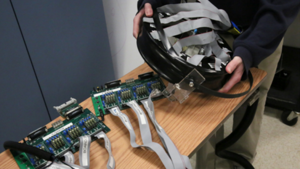
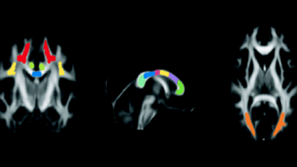
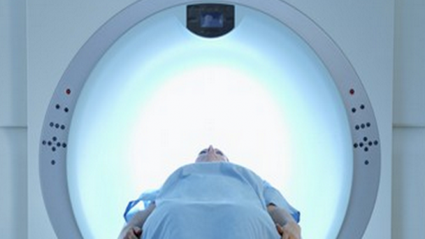
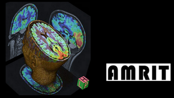
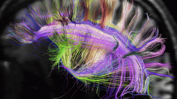
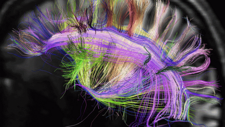
The Martinos Center for Biomedical Imaging dual mission includes translational research using state-of-the-art imaging technologies and ongoing development of those technologies. The core technologies being developed and used at the Center include magnetic resonance imaging (MRI), positron emission tomography (PET) and more (see the navigation menu on the left for a complete list of the technologies). A key area of innovation is Multimodal Functional Neuroimaging, which involves the integration of two or more different imaging technologies.

Multimodal Functional Neuroimaging, which involves the integration of two or more different imaging technologies.

Web Information

Website:  www.martinos.org/ Brain Initiative Grant

Contact Information

Email: info@martinos.org Phone: 617-726-3197 Address: Athinoula A. Martinos Center for Biomedical Imaging 149 Thirteenth Street, Suite 2301 Charlestown, Massachusetts 02129

About

The Athinoula A. Martinos Center for Biomedical Imaging at Massachusetts General Hospital is one of the world’s premier research centers devoted to development and application of advanced biomedical imaging technologies. Our mission is to advance imaging in healthcare through technology development, translational research and education.

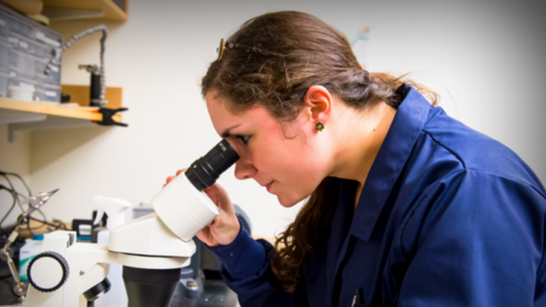
Located on the MGH Research Campus in Charlestown, the Center is home to roughly 100 faculty researchers and more than 200 affiliated and visiting faculty, postdoctoral research fellows and graduate students, who use advanced imaging technologies both separately ...

OnAir Post: Center for Biomedical Imaging

Skip to toolbar