Calcium sensors for molecular fMRI

PI: Alan Jasanoff Massachusetts Institute of Technology Title: “Calcium sensors for molecular fMRI” BRAIN category: Large-Scale Recording-Modulation – New Technologies (RFA NS-14-007)

Dr. Jasanoff’s team will synthesize calcium-sensing contrast agents that will allow functional magnetic resonance imaging (fMRI) scans to reveal activity of individual brain cells.

NIH Webpages

Project Description

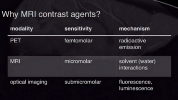
The development of minimally invasive direct readouts of neural activity is one of the greatest challenges facing neuroscience today. Our recent work has shown that it is possible to perform high resolution functional magnetic resonance imaging (fMRI) of molecular-level phenomena using MRI contrast agents sensitive to hallmarks of neurotransmitter release. An even more valuable contribution would be the creation of calcium sensors suitable for molecular fMRI of intracellular neural signaling processes. Functional imaging performed with these sensors would combine the noninvasiveness and whole-brain coverage of MRI with the molecular specificity and broad applicability of established optical calcium neuroimaging techniques. Calcium-dependent fMRI will be a breakthrough technique for analysis of neural circuits in animals, with potential longer term applications in humans. The technique could achieve cellular resolution in conjunction with ultrahigh field MRI scanners and cell labeling techniques. A major hurdle in realizing this advance is the creation of effective calcium-dependent MRI contrast agents, however. This proposal describes ...

OnAir Post: Calcium sensors for molecular fMRI

High-Density Recording Microelectrodes

PI: Tim Gardner, Laboratory of neural circuit formation Institution: Boston University (Charles River Campus) Title: “High-Density Recording and Stimulating Microelectrodes” BRAIN Category: Large-Scale Recording-Modulation – New Technologies (RFA NS-14-007)

Dr. Gardner and his colleagues will develop ultrathin electrodes that minimize tissue damage and are designed for long-term recording of neural electrical activity.

NIH Webpages

Time-frequency microstructure is unstable for regions of the time-frequency plane with spectrally dense content. For those regions, small changes in analysis parameters or added background noise in the signal can lead to changes in the details of a sonogram or contour shapes. Structurally unstable portions of the representation can be eliminated by showing only contour fragments that are in agreement across different angles and time-scales of analysis.The image below illustrates that process. On top, all long contours are shown, weighted by sonogram power.On bottom, only the structurally stable “consensus” elements are shown, also weighted by sonogram power.

Project Description

This project seeks to develop a high density, minimally invasive electrode array for long-term recording and control of brain activity. Multielectrode arrays are an essential tool in experimental and clinical neuroscience, yet current arrays are severely limited by a mismatch between large or stiff electrodes and the fragile environment ...

OnAir Post: High-Density Recording Microelectrodes

Modular nanophotonic probes

Principal Investigator: Michael Roukes Caltech Neuroscience Title: “Modular nanophotonic probes for dense neural recording at single-cell resolution” BRAIN Category: Large-Scale Recording-Modulation – New Technologies (RFA NS-14-007)

Dr. Roukes and his team propose to build ultra-dense, light-emitting and -sensing probes for optogenetics, which could simultaneously record the electrical activity of thousands of neurons in any given region of the brain.

NIH Webpages

Nanoprobes, Wireless, and Synthetic Biology Technologies for the BAM Project(Left) Silicon nanoprobe arrays (after Du et al., 2009b ). (A) Flip-chip assembly scheme for connecting the silicon devices with printed circuit boards. (B) SEM micrograph of the rear section of a 50-μm-thick shaft array showing the multilayer stacked structure. Adjacent layers have a spacing of 100 μm, which is set by the thickness of the flexible cable. (C) Side view of the 50-μm-thick shaft array showing that the shafts are stress balanced and are able to retain approximately constant relative spacing.(Right) Synthetic biology approaches. (D) A voltage sensitive calcium channel influences the error rate of an engineered DNA polymerase. X marks sites of mismatch between “T” in the template strand (lower) and “G” new copy strand. Note scale of the various devices and cells.

Project Description

Our understanding of ...

OnAir Post: Modular nanophotonic probes

Diamond Electrodes for Measurement

Principal Investigator: Kendall H  Lee Mayo Clinic Rochester Title: “Neurotransmitter Absolute Concentration Determination with Diamond Electrode” BRAIN Category: Large-Scale Recording-Modulation – New Technologies (RFA NS-14-007)

Dr. Kendall and his colleagues will develop diamond-coated electrodes to measure concentrations of the brain chemical dopamine more accurately and over long periods of time in the brain.

NIH Webpages

An implantable diamond-coated flexible electrode. Image from Qmed.

Project Description

Determining the levels of neurotransmitters present in the living brain in real time is a matter of current scientific interest for research and clinicl reasons. Among these reasons is the need for understanding and mapping brain function and for improvement in the clinical application of deep brain stimulation (DBS). One analytical technique that holds potential promise in this application is fast-scan cyclic voltammetry (FSCV), however technical limitations have hindered its adoption for chronic in vivo use. Of particular difficulty has been the construction of a chronically-implantable FSCV electrode that possesses both the proper chemical properties for the monitoring of neurotransmitter levels as well as sufficient durability for chronic implantation in humans or animals. Carbon fiber has been used successfully under some circumstances, particularly at low voltage potentials, but at the higher voltages required for detection ...

OnAir Post: Diamond Electrodes for Measurement

Genetic sensors for biogenic amines

Principal Investigator: Lin Tian UC Davis Neuroscience Title: ” Genetically encoded sensors for the biogenic amines: watching neuromodulation in action” BRAIN Category: Large-Scale Recording-Modulation – New Technologies (RFA NS-14-007)

Dr. Tian and her colleagues will create sensors that will allow researchers to see how molecules like dopamine, norepinephrine and serotonin regulate activity of neural circuits and behavior in living animals.

NIH Webpages

Genetically encoded indicators of neural activity.Fluorescent protein based biosensors can transfer changes in neural state (e.g. membrane potential or essential ion flux or enzyme activity) to fluorescence observables. They are genetically encoded, and can thus be used to label large populations of defined cell types and/or sub-cellular compartments.

Project Description

The goal of this proposal is to develop a toolbox of genetically encoded indicators for biogenic amines, the most important family of neuromodulators. All nervous systems are subject to neuromodulation, which reconfigure the dynamics of neural circuitry by transforming the intrinsic firing properties of targeted neurons and regulating their synaptic plasticity. The altered dynamics of the neuromodulators have been implicated in a number of human neurological and psychiatric diseases, including Parkinson’s, schizophrenia and addiction. Biogenic amines are a group of neuromodulators used by all animal brains to regulate the ...

OnAir Post: Genetic sensors for biogenic amines

Optogenetic mapping of synaptic activity

Principal Investigator: John Yu-Luen Lin Neuroscience at UCSD Title: “Optogenetic mapping of synaptic activity and control of intracellular signaling” BRAIN Category: Large-Scale Recording-Modulation – New Technologies (RFA NS-14-007)

Dr. Lin’s team will create molecules that, when they are triggered by a pulse of light, allow scientists to test for communication between neurons in specific circuits of the brain.

NIH Webpages

Project Description

This proposal aims to develop new molecular techniques to map activities of neurons, manipulate the strength of communication between neurons and disrupt intracellular signaling. These ‘optogenetic’ approaches will be used to further our understandings of brain function on behavior and have important implications in our understandings of neurological conditions and neurodegenerative diseases. The first goal is to develop a technique where the researchers can use optical approach to identify synaptic connections that were active during the performance of a behavior task. This reporter system can be turned on with light, which defines the window of activity reporting, and fluorescence signal can be detected if there is significant activity between two defined cell groups. Many existing approaches can only be used to map excitatory connections, whereas the proposed approach can be used to identify activities between synapses utilizing any neurotransmitters. The approach will utilize ...

OnAir Post: Optogenetic mapping of synaptic activity

Modular systems measuring brain activity

Principal Investigator: Loren Frank UCSF Neuroscience Title: ” Modular systems for measuring and manipulating brain activity” BRAIN Category: Large-Scale Recording-Modulation – New Technologies (RFA NS-14-007)

Dr. Frank and his colleagues will engineer a next-generation, all-in-one neural recording and stimulating system, which can simultaneously monitor thousands of neurons in the brain for several months while also delivering drugs, light or electrical pulses.

NIH Webpages

NSpike Instructions – The diagram shows the overall organization of the data acquisition system as it is used in the Frank lab,

Project Description

The brain is a massively interconnected network of specialized circuits. Even primary sensory areas, once thought to support relatively simple, feed-forward processing, are now known to be parts of complex feedback circuits. All brain functions depend on millisecond timescale interactions across these brain networks, but current approaches cannot measure or manipulate these interactions with sufficient resolution to resolve them. We need the capacity to measure and manipulate the activity large ensembles of neurons distributed across anatomically or functionally connected circuits. That technology does not yet exist, a lack that motivates our efforts to develop a new system for large scale, multisite recording and manipulation that takes integrates biocompatible polymer electrodes, new headstage ...

OnAir Post: Modular systems measuring brain activity

Optoelectrodes for Local Circuit Analysis

Principal Investigator: Euisik  Yoon University of Michigan Neuroscience  Title: ” Modular High-Density Optoelectrodes for Local Circuit Analysis” BRAIN Category: Large-Scale Recording-Modulation – New Technologies (RFA NS-14-007)

In this project, Dr. Yoon’s team will make devices for optogenetics, a technique that enables scientists to turn neurons on and off with flashes of light, more precise and diverse by integrating multiple light sources in such a way as to enable the control of specific neuronal circuits.

NIH Webpages

Analog Front-End Module With Moderate Inversion and Power-Scalable Sampling Operation for 3-D Neural MicrosystemsWe report an analog front-end prototype designed in 0.25 CMOS process for hybrid integration into 3-D neural recording microsystems. For scaling towards massive parallel neural recording, the prototype has investigated some critical circuit challenges in power, area, interface, and modularity.

Project Description

A number of scientific questions, especially in local circuit analysis, require manipulating neurons in vivo at multiple sites independently at high spatial and temporal resolutions by perturbing a controlled number and simultaneously recorded neurons. Optogenetic stimulation is cell-type specific which has proven to be the most powerful means of circuit control. Several laboratories have developed solutions to deliver optical stimulation to deep brain structures whilst simultaneously recording neurons. However, stimulation ...

OnAir Post: Optoelectrodes for Local Circuit Analysis

Deep Photoacoustic Tomography

Principal Investigator: Lihong Wang Washington University Neuroscience Program Title: “Fast High-Resolution Deep Photoacoustic Tomography of Action Potentials in Brains” BRAIN Category: Large-Scale Recording-Modulation – New Technologies (RFA NS-14-007)

Dr. Wang and his collaborators will test a way to image the electrical activity of neurons deep inside the brain, using a variation on ultrasound imaging he invented called photoacoustic tomography.

NIH Webpages

https://www.youtube.com/watch?v=yNPqOuk_YdgVideo can’t be loaded because JavaScript is disabled: Lihong Wang Hot Topics presentation: Photon-Phonon Synergy: Photoacoustic Tomography and Beyond (https://www.youtube.com/watch?v=yNPqOuk_Ydg)

Project Description

Revealing how our brain works is a great challenge but yet worth our every effort: it will not only illuminate the profound mysteries in science but also provide the key to understanding and treating neurological diseases such as Alzheimer’s and Parkinson’s. The objective of the proposed three-year research is to develop a high- speed, high-spatial-resolution, deep-penetration photoacoustic computed tomography (PACT) system for real- time imaging of action potentials in mouse brains. The proposed hardware imaging system will be unprecedented in the field of PACT in ...

OnAir Post: Deep Photoacoustic Tomography

Genetically encoded reporters

Principal Investigator: Kit S. Lam UC Davis Center for Neuroscience Title: “Genetically encoded reporters of integrated neural activity for functional mapping of neural circuitry” BRAIN Category: Large-Scale Recording-Modulation – New Technologies (RFA NS-14-007)

Dr. Lam’s team plans to develop fluorescent sensors that will mark ion channels, molecules that help control information flow in the brain, and enable scientists to observe the neurons that are activated during a specific behavior, such as running.

NIH Webpages

Graphic from Shultz Chemical Cell Biology Group at Heidelberg

Project Description

One of the major challenges in neuroscience is to link the structure to the function of neural circuits. To achieve this goal, we need to understand the connectivity between defined neuronal populations and the contribution of these neurons to physiological processes, behavioral responses and disease states. Recent advances in imaging techniques allow us to visualize the brain structure with cellular resolution. Application of the current generation of genetically encoded optical tools, such as sensors and controllers, is facilitating measurement and manipulation of neuron activity from molecular-defined cell populations in awake, behaving animals. However, probing the dynamics of neural circuitry underlying behavior, specifically for dissecting functional-defined circuitry beyond molecular-defined circuitry, not only depends on the improvement ...

OnAir Post: Genetically encoded reporters

Time-Reversal Optical Focusing

Principal Investigator: Changhuei Yang Caltech Neuroscience Title: Time-Reversal Optical Focusing for Noninvasive Optogenetics BRAIN Category:  Large-Scale Recording-Modulation – New Technologies (RFA NS-14-007)

Dr. Yang’s team plans to develop a light and sound system that will noninvasively shine lasers on individual cells deep within the brain and activate light-sensitive molecules to precisely guide neuronal firing.

NIH Webpages

Optofluidic microscopy (OFM) is a new compact and lensless microscopic imaging technique invented in our lab. It abandons the conventional microscope design, which requires expensive lenses and large space to magnify images, and instead utilizes microfluidic flow to deliver specimens across array(s) of micrometer-size apertures defined on a metal-coated CMOS sensor to generate direct projection images. The size of our OFM prototype device is as small as a US quarter, and yet can render images comparable in quality to those of a microscope with 20X objective.

Project Description

Our bodies appear optically opaque because biological tissue scatters light strongly. Although advances such as multiphoton excitation have enabled deeper access for optical imaging by gating out scattered light, these strategies are still fundamentally limited to superficial depths (~ 1 mm). Yang’s group at Caltech has pioneered time-reversal symmetry of optical scattering as a direct strategy ...

OnAir Post: Time-Reversal Optical Focusing

Skip to toolbar