Ecker Lab – Salk

Principal Investigator: Joseph R Ecker Salk Institute for Biological Studies

Being able to study the epigenome in great detail and in its entirety will provide a better understanding of plant productivity and stress resistance, the dynamics of the human genome, stem cells’ capacity to self-renew and how epigenetic factors contribute to the development of tumors and disease. We are now exploring how DNA methylation effects the development of human embryonic stem (hES) cells as well as induced pluripotent stem (IPS) cells as they are induced to differentiate into other types of cells.


Web Information

Website:  pbio.salk.edu/pbioe/members Brain Initiative Grant

Contact Information

Phone: (858) 453-4100 x1752 Address: Salk Institute Genomic Analysis Laboratory 10010 N. Torrey Pines Road La Jolla, CA 92037

Research

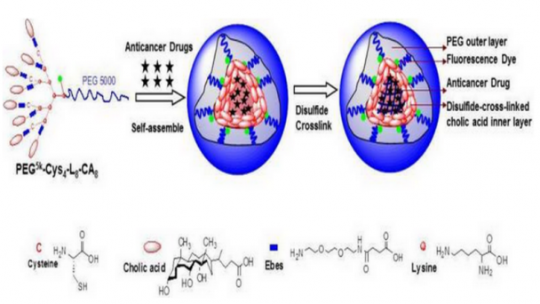
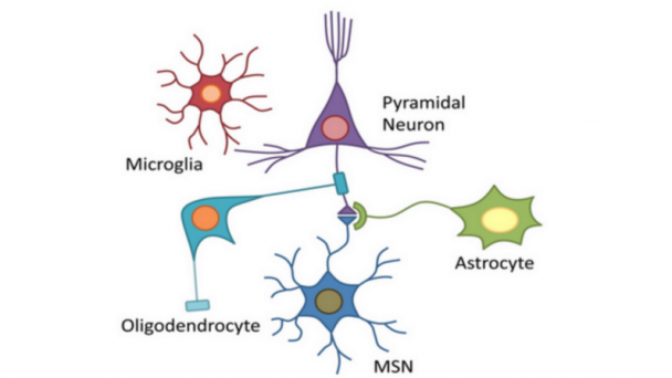
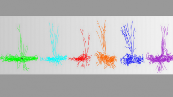
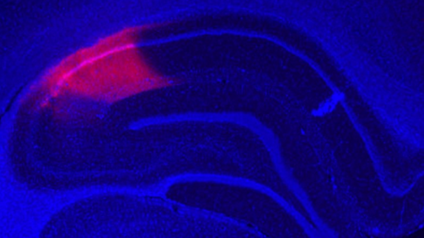
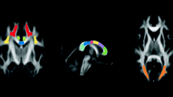
The development of DNA sequencing technologies that produce vast amounts of sequence information has triggered a paradigm shift in biology, enabling massively parallel surveying of complex nucleic acid populations. The diversity of applications to which these technologies have already been applied demonstrates the immense range of cellular processes and properties that can now be studied at the single-base resolution. These applications include, but are not limited to, the sequencing of genomes to uncover nucleotide polymorphisms and structural variation, as well as epigenomes to reveal sites of DNA–protein interaction and ...

OnAir Post: Ecker Lab – Salk

Precision Biosystems Laboratory – Ga. Tech

The Precision Biosystems Laboratory is focused on fundamental engineering advancements, the development of miniaturized, high-throughput robotic instrumentation, and the application of the two to advance biomolecular science. Their research program is in the emerging bio-nano field—at the intersection of bioMEMS, machine design, neuroengineering, genetics, optics, and manufacturing.

OnAir Post: Precision Biosystems Laboratory – Ga. Tech

Roth Lab – UNC

Principal Investigator: Bryan Roth UNC Neuroscience

Roth Lab studies the structure and function of G-Protein coupled receptors (GPCRs). Roth Lab is part of the Department of Pharmacology, a research department in the School of Medicine at the University of North Carolina at Chapel Hill. The Roth lab is the principal contractor for the NIMH Psychoactive Drug Screening Program which includes the PDSP Ki database.

Serotonin receptor neurobiology: Since 1984, my lab has studied serotonin (5-hydroxytryptamine; 5-HT) and its receptors. Recently we have been focused on targeting and trafficking of 5-HT2-family receptors . Ongoing projects are directed to using mouse genetics to delineate the roles of accessory proteins and post-translational modifications in 5-HT receptor actions.

Web Information

Website:  pdspdb.unc.edu/rothlab/ BRAIN Initiative Grant – ” Dreadd2.0: An Enhanced Chemogenetic Toolkit”

Contact Information

Email: estelalopez@unc.edu Phone: 919-966-7535 Address: 4072 Genetic Medicine UNC-CH School of Medicine Chapel Hill, NC 27599-7365

Research

The Roth lab perfected the chemogenetic technology we have named “DREADD” (Designer Receptor Exclusively Activated by Designer Drugs; Armbruster et al, 2007). DREADD technology has afforded 100’s of labs world-wide the opportunity to discover how cell-type specific modulation of signaling is translated into behavioral and non-behavioral outcomes (see Urban and Roth, Ann Rev Pharmacol Toxicol 2015 for recent review)

The Roth lab continues to enhance ...

OnAir Post: Roth Lab – UNC

Schnitzer Group – Stanford

Principal Investigator: Mark J Schnitzer Stanford Neurosciencs Institute

The Schnitzer Group has three major research efforts: Development and application of fiber-optic, micro-optic, and nanophotonic imaging techniques for studies of learning and memory in behaving mice and for clinical uses in humans; In vivo fluorescence imaging and behavioral studies of hippocampal-dependent cognition and learning; and Development of high-throughput, massively parallel imaging techniques for studying brain function in large numbers of Drosophila concurrently.


Web Information

Website:  http://pyramidal.stanford.edu/index.html Brain Initiative Grant

Contact Information

Email: schnizerlab@gmail.com Address: James H. Clark Center – Room W080 318 Campus Drive Stanford, CA 94305

Research

Dr. Schnitzer has longstanding interests in neural circuit dynamics and optical imaging, and his laboratory has three major research efforts:

In vivo fluorescence imaging and behavioral studies of cerebellar-dependent motor control and motor learning. Development and application of fiber-optic fluorescence microendoscopy imaging techniques for studies of learning and memory in behaving mice and for clinical uses in humans. Development of high-throughput, massively parallel imaging techniques for studying brain function in large numbers of Drosophila concurrently.

The long-term goal of our research is to advance experimental paradigms for understanding normal cognitive and disease processes at the level of neural circuits, with emphasis on learning and memory processes. By contrast, much current research on learning and memory concentrates on ...

OnAir Post: Schnitzer Group – Stanford

IDV – The Vision Institute

Research Director: Serge Picaud Pierre and Marie Curie University

Built in the heart of the Quinze-Vingts National Eye Hospital, the Vision Institute is one of the most important research centers in Europe on eye diseases.The Institute’s goal is to discover, test and develop treatments and technological innovations of tomorrow in order to prevent or limit visual impairment and to improve the autonomy and the quality of life of patients.

Web Information

Website:  institut-vision.org/ Brain Initiative Grant

Contact Information

Email: contact@institut-vision.org Phone: 33 1 53 46 26 48 Address: The Vision Institute 17 rue Moreau 75012 Paris – France

OnAir Post: IDV – The Vision Institute

Laboratory of Neural Circuit Dynamics – Zurich

To study neural circuit function our research is focused on advancing and applying in vivo high-resolution imaging methods, with a particular emphasis on neocortical microcircuitry. The lab's specific goals are to reveal principles of single-cell and local network computation and to decipher the neural codes governing information processing as well as circuit plasticity.

OnAir Post: Laboratory of Neural Circuit Dynamics – Zurich

Xu Research Group – Cornell

Principal Investigator, Chris Xu Cornell University

Xu Research Group has two main thrusts: biomedical imaging and fiber optics. The Group is exploring new concepts and techniques for in vivo imaging deep into scattering biological specimens, such as mouse brain; developing new medical endoscopes for non-invasive real-time diagnostics of tissues without any exogenous contrast agent and novel optical fibers and fiber-based devices for biomedical imaging and optical communications.

Nonlinear Optics in Higher-order-mode Fibers. Xu Research Group

Web Information

Website:  http://xu.research.engineering.cornell.edu/ Brain Initiative Grant

Contact Information

EmailCX10@cornell.edu Phone: 607 255-1460 Address: 276 Clark Hall:

Research

    Our research has two main thrusts: biomedical imaging and fiber optics. We are exploring new concepts and techniques for in vivo imaging deep into scattering biological specimens, such as mouse brain. We are developing new medical endoscopes for non-invasive real-time diagnostics of tissues without any exogenous contrast agent. We are developing novel optical fibers and fiber-based devices for biomedical imaging and optical communications.

Biomedical Imaging

Deep Imaging in Scattering Biological Tissue Laser scanning multiphoton microscopy (MPM) has greatly improved the penetration depth of optical imaging and is proven to be well suited for a variety of imaging applications deep within intact or semi-intact tissues. Nonetheless, MPM ...

OnAir Post: Xu Research Group – Cornell

Kanold Lab – UMD

Principal Investigator: Patrick Kanold UMD Neuroscience and Cognitive Science

The major focus in the lab is to understand how information about the world is represented/encoded in the brain, the circuits present in the adult and developing brain and their influence of brain development and plasticity. One focus is on probing the response of the brain to sensory stimuli and the other is to record from small sub-circuits and study their responses and circuit behavior in great detail. 


Web Information

Website:  clfs.umd.edu/biology/kanold BRAIN Initiative Grant – “Crowd coding in the brain: 3D imaging and control of collective neuronal dynamics”

Contact Information

Emailpkanold@umd.edu Phone: 301.405.5741 Address: 1116 Bioscience Research Building College Park, MD 2074

Research

How do our brains work, wire up, and change?

      The human brain is a tremendously complex neuronal circuit that we don’t really understand. For example we do not know how sensory information about the world is encoded in neuronal population activity and transformed into a percept.

    Moreover, somehow during development all the highly specific connections in the brain are made so that we can function normally. It is clear now that we do not come hard-wired but that interaction with the environment plays a major role in shaping our brain. In particular during

OnAir Post: Kanold Lab – UMD

Grossman Institute – Chicago

Principal Investigator: John Maunsell Neuroscience at University of Chicago

Grossman Institute for Neuroscience, Quantitative Biology and Human Behavior is a new Institute designed to establish a group of scholars working at the intersection of quantitative biology, neuroscience, and the study of social and individual behaviors. The Institute will build upon existing strengths in these fields to address fundamental questions about the biological, social, and environmental factors that shape social behaviors and inter-individual variation in model organisms and humans.


Web Information

Website: uchicago.edu/grossman_institute_for_neuroscience_quantitative_biology_and_human_behavior BRAIN Initiative Grant – “The role of patterned activity in neuronal codes for behavior”

Contact Information

Phone: 773.702.1234

Address: The University of Chicago Edward H. Levi Hall 5801 South Ellis Avenue Chicago, Illinois 60637

Research

The Grossman Institute for Neuroscience, Quantitative Biology, and Human Behavior is the intellectual home for a diverse group of scholars and scientists working together to advance our understanding of the brain. By utilizing a multidisciplinary approach, the Institute will recruit faculty members from such areas as Chemistry, Cognitive Sciences, Computer Science, Genetics, Molecular Biology, Statistics and many more to reveal how the human brain and our social and physical environments interact.

Articles

Inquiring minds

University of Chicago Magazine Nov-Dec. 2014 by Kevin Jiang

Neuroscientist John Maunsell leads a new institute’s research into ...

OnAir Post: Grossman Institute – Chicago

David Kleinfeld Laboratory – UCSD

Principal Investigator: David Kleinfeld UCSD Neuroscience;  Neurophysics Research

Active sensation: how orofacial behaviors, with emphasis on the vibrissa sensorimotor system, encode a stable world view through actively moving sensors. Microcirculation in the Brain: the structure and control of cortical blood flow, and variations in that flow, at the level of vascular networks down to that of individual microvessels. CNiFERs: uses of cell-based sensors of signaling molecules to study volume transmission and neuromodulation in behaving animals.


Web Information

Website:  physics.ucsd.edu/neurophysics/index.php BRAIN Initiative Grant – “Revealing the connectivity and functionality of brain stem circuits”

Contact Information

Email: dk@physics.ucsd.edu Phone: 858-822-0342 Address: Physics Department UC San Diego

Research

Active sensation:

This program addresses how orofacial behaviors, with emphasis on the vibrissa sensorimotor system, encode a stable world view through actively moving sensors.

Microcirculation in the Brain:

This program explores the structure and control of cortical blood flow, and variations in that flow, at the level of vascular networks down to that of individual microvessels.

CNiFERs:

This program creates cell-based sensors of signaling molecules to study volume transmission and neuromodulation in the brains of behaving animals.

Publications

OnAir Post: David Kleinfeld Laboratory – UCSD

Nelson Lab – Brandeis

Principal Investigator: Sacha B. Nelson Brandeis University

Despite their functional and clinical importance, the cell types that comprise the neocortex and the molecular mechanisms that specify their properties and connectivity are only partly understood. Nelson Lab studies the development and function of the neocortex in the laboratory mouse using a combination of genetic, genomic and electrophysiological approaches.

In our approach to examining the neocortex we use new driver strains developed here and by our collaborators to genetically or virally deliver mutant alleles to specific neuronal cell types. We monitor effects on physiology and connectivity using patch clamp recording and high resolution anatomy. To evaluate changes in gene expression we developed methods for manually sorting fluorescent neurons and performing genome-wide expression profiling. Nelson Lab

Web Information

Website: bio.brandeis.edu/nelsonlab/ Brain Initiative Grant

Contact Information

Email: nelson@brandeis.ed Phone: (781) 736-3181 Address: Brandeis University 415 South Street, Waltham, MA 02454 Shapiro Science Center Room 1-15

Research

The Nelson Lab is located in the recently constructed Shapiro Science Center at Brandeis University located in Waltham, Massachusetts and is a member of the National Center for Behavioral Genomics. We are primarily interested in examining the mammalian neocortex. Our lab combines electrophysiology, advanced imaging techniques, mouse genetics and high throughput gene expression analysis allowing us ...

OnAir Post: Nelson Lab – Brandeis

Sestan Lab – Yale

Principal Investigator: Nenad Sestan Yale Neuroscience

The Sestan Lab’s research centers on understanding the molecular and cellular basis of how neurons acquire distinct identities and form proper synaptic connections in the cerebral cortex, a part of the brain that is critical for cognition, perception and behavior. The Lab also studies how these complex developmental processes have evolved and become compromised in human disorders, such as autism. An important element of our research is the integration of complementary approaches.


Web Information

Website:  medicine.yale.edu/lab/sestan/ BRAIN Initiative Grant – “A Novel Approach for Cell-Type Classification and Connectivity in the Human Brain”

Contact Information

Email: sestanlab@yale.edu Phone: 203.737.1435 Address: Sterling Hall of Medicine 333 Cedar Street, SHM C-316C New Haven, CT 06510

Research Interests

Our research centers on understanding the molecular and cellular basis of how neurons acquire distinct identities and form proper synaptic connections in the cerebral cortex, a part of the brain that is critical for cognition, perception and behavior. We also study how these complex developmental processes have evolved and become compromised in human disorders, such as autism. An important element of our research is the integration of complementary approaches that combines 1) analyses of evolutionarily conserved developmental mechanisms using the genetically tractable mouse model, 2) comparative genomic and cellular analyses of non-human primates and humans to identify human-specific features of ...

OnAir Post: Sestan Lab – Yale

Kit Lamb Lab – UCDavis

The main focus of the Lam Lab lies in discovering revolutionary and innovative methods of disease treatment on a nano-scale and molecular scale. The lab work is most related to molecular medicine, drug discovery, nanoparticle and drug-delivery techniques in cancer and other diseases. Our research is done through the use of cutting edge technologies and established procedures.

OnAir Post: Kit Lamb Lab – UCDavis

Optical Imaging Laboratory – Washington U

Optical Imaging Laboratory develops novel biophotonic tomography for early-cancer detection and functional imaging, using non-ionizing electromagnetic and ultrasonic waves. Research directions include: Photo-acoustic tomography (PAT), Thermo-acoustic tomography (TAT), Ultrasound-modulated (acousto-) optical tomography (UOT), Mueller optical-coherence tomography (M-OCT), Oblique-incidence reflectometry (OIR) and spectroscopy, and Modeling light transport in tissues.

OnAir Post: Optical Imaging Laboratory – Washington U

Yoon Lab – Michigan

Principal Investigator: Euisik  Yoon University of Michigan Neuroscience

The mission of the Yoon lab is to build self-contained microsystems that will combine and process natural signals (such as bio, chemical, optical and thermal signals) as well as electrical signals on a single chip platform by integrating new MEMS/nano structures with low-power, wireless VLSI circuits and systems. Currently, we have three main research thrusts: microfluidic systems, MEMS neural interfaces, and CMOS imaging and neural interfacing ICs.

Web Information

Website:  yoon.eecs.umich.edu/ BRAIN Initiative Grant – ” Modular High-Density Optoelectrodes for Local Circuit Analysis”

Contact Information

Email: esyoon@umich.edu Address: 1301 Beal Avenue, Ann Arbor

Research

The mission of our lab is to build self-contained microsystems that will combine and process natural signals (such as bio, chemical, optical and thermal signals) as well as electrical signals on a single chip platform by integrating new MEMS/nano structures with low-power, wireless VLSI circuits and systems. Currently, we have three main research thrusts: microfluidic systems, MEMS neural interfaces, and CMOS imaging and neural interfacing ICs.


Advanced Neural Probes

Mapping the brain and peripheral circuits may be the grandest challenge in a all of science today. Our mission is to help neuroscientists, neurologists, and other clinicians understand these amazingly complex circuits with tools that can monitor neural activity or recreate ...

OnAir Post: Yoon Lab – Michigan

Brefczynski-Lewis Lab – WVU

Brefczynski-Lewis Lab studies how we perceive people we love and people we don’t like, both famous and political, and how training in compassion can affect those perceptions. The Lab is examining the neural and physiological correlates of the liked and disliked persons and how these change after training in compassion. Grudge forgiveness study: fMRI response to the face of the grudge person, as well as cardio and reactive measures will be tested before and after the intervention.

OnAir Post: Brefczynski-Lewis Lab – WVU

Laboratory of Molecular Genetics – Rockefeller

Principal Investigator: Jeffrey M. Friedman Senior Research Associate: Sarah Stanley, Rockefeller University

The application of modern methods in genetics has led to the identification of a new hormone, leptin, that regulates body weight. Leptin is an adipose tissue hormone that interacts with receptors in the brain to regulate food intake, energy expenditure and other neuroendocrine systems. The molecular mechanisms of leptin in the brain are under investigation. These studies are being conducted in parallel with efforts to identify obesity genes in the human.

Diagram showing the phosphorylation sites on ribosomal protein S6 and strategy for immunoprecipitation of phosphorylated ribosomes. from A critical role for mTORC1 in erythropoiesis and anemia.

Web Information

Website:  lab.rockefeller.edu/friedman/ Brain Initiative Grant

Contact Information

Email: Jeffrey.Friedman@rockefeller.edu Phone: (212) 327-8000 Address: The Rockefeller University 1230 York Avenue New York, NY 10065 (212) 327-8000

Research

The application of modern methods in genetics has led to the identification of a new hormone, leptin, that regulates body weight. Leptin is an adipose tissue hormone that interacts with receptors in the brain to regulate food intake, energy expenditure and other neuroendocrine systems. The molecular mechanisms of leptin in the brain are under investigation. These studies are being conducted in parallel with efforts to identify obesity genes in the human.

Research Projects:

OnAir Post: Laboratory of Molecular Genetics – Rockefeller

Rubenstein Lab – UCSF

Principal Investigator: John L. R. Rubenstein UCSF Neuroscience

The goal of Rubenstein Lab research is to elucidate fundamental mechanisms that regulate development of the forebrain, with a focus on the cerebral cortex and basal ganglia. Our studies also extend into other regions of the embryo, including the developing face. Whenever possible, we attempt to investigate whether disruption of these mechanisms underlie human disorders, such as autism, schizophrenia, mental retardation, epilepsy and craniofacial disorders.


Web Information

Website:  rubensteinlab.ucsf.edu/ BRAIN Initiative Grant – “Identification of enhancers whose activity defines cortical interneuron types”

Contact Information

Email: john.rubenstein@ucsf.edu Phone: 415-476-7862 Address: John L.R. Rubenstein, M.D., Ph.D. Genetics, Development and Behavioral Sciences Building 1550 4th Street, 2nd Floor South, Room GD 284C University of California at San Francisco San Francisco, CA 94143-2611

About the Lab

Mission The goal of our research is to elucidate fundamental mechanisms that regulate development of the forebrain, with a focus on the cerebral cortex and basal ganglia. Our studies also extend into other regions of the embryo, including the developing face. Whenever possible, we attempt to investigate whether disruption of these mechanisms underlie human disorders, such as autism, schizophrenia, mental retardation, epilepsy and craniofacial disorders. We hope that our studies provide insights into new inroads for diagnosis, prevention and treatment of these disorders.

History Our research group began at Stanford in 1988 to ...

OnAir Post: Rubenstein Lab – UCSF

X. William Yang Research Group – UCLA

Yang Lab's research is focused on applying comprehensive molecular and genetic approaches to study the pathogenesis of Huntington’s disease (HD) and Parkinson’s disease (PD), as well as exploring the molecular genetics and circuitry of the basal ganglia (BG), a brain region targeted in various neurodegenerative and neuropsychiatric disorders. The overarching approach of our lab is to develop genetic mouse models of these diseases, or create mice that carry mutations in the genes.

OnAir Post: X. William Yang Research Group – UCLA

Scanziani Lab – UCSD

Principal Investigator: Massimo Scanziani UC San Diego’s Neuroscience

The goal of Scanziani Lab’s research is to understand the mechanisms by which elementary circuits of neurons control the spatial and temporal structure of cortical activity. Towards this goal they use in vivo and in vitro electrophysiological, imaging and anatomical techniques as well as behavioral approaches. Their model system is the rodent’s sensory cortex.

Web Information

Website:  scanzianilab.org/ Brain Initiative Grant – “Classifying Cortical Neurons by Correlating Transcriptome with Function”

Contact Information

Email: massimo@ucsd.edu Phone: (858) 822-3840 Address: Center for Neural Circuits and Behavior, Room 213 9500 Gilman Dr. La Jolla CA 92093-0634

Research

Sensations and thoughts result from the coordinated activity of neuronal populations in space and time. The goal of my research is to understand the mechanisms by which elementary circuits of neurons control the spatial and temporal structure of cortical activity. Towards this goal we use in vivo and in vitro electrophysiological, imaging and anatomical techniques as well as behavioral approaches. Our model system is the rodent’s sensory cortex.

Our work is revealing the logic and the mechanisms by which elementary circuits, the building blocks of cortical architecture, orchestrate cortical activity.

Publications

Visual Cortex

Xue M, Atallah BV, Scanziani M.

Equalizing exitation-inhibition ratios across visual cortical neurons.

Nature 511: 596-600 (2014) [pdf]

Bortone DS, Olsen SR, ...

OnAir Post: Scanziani Lab – UCSD

Laboratory of neural circuit formation – BU

Principal Investigator, Tim Gardner
Boston University (Charles River Campus)

The Gardner lab studies the mechanisms of temporal sequence perception and production, focusing on vocal learning in songbirds. The Laboratory of neural circuit formation current research projects involve: Sensory-motor learning; High-density recording and stimulating microelectrodes, Electrode arrays for the central nervous system: tissue interaction; and Peripheral nervous system : chronic recording and stimulation for biolectric medicine.

OnAir Post: Laboratory of neural circuit formation – BU

Mayo Physiology and Biomedical Engineering Dept.

The Department of Physiology and Biomedical Engineering at Mayo has a long and rich history of scientists and physicians collaborating and translating basic discoveries. The department currently consists of 18 primary and 35 joint appointees. There are 34 doctoral students, 67 research fellows and research associates working in a variety of research areas.

OnAir Post: Mayo Physiology and Biomedical Engineering Dept.

Soltesz Lab – UCIrvine

The Soltesz Lab is interested in how brain cells communicate with each other and how the communication changes after fever-induced seizures in early childhood and after head injury. Our general goal is to understand how neuronal networks function and dysfunction, in order to discover new therapies to prevent epilepsy.

OnAir Post: Soltesz Lab – UCIrvine

Kramer Lab – Stanford

Principal Investigator: Richard  Kramer UC Berkeley Helen Wills Neuroscience Institute

Kramer Lab studies utilize novel chemical reagents to modify the function of ion channels and synapses.  This Chemical-Biological approach is designed to allow non-invasive optical sensing and optical manipulation of channels and synapses in the nervous system. One major goal of this research is to develop the technology for restoring vision in degenerative blinding diseases.

Regulating native ion channels with light – we have sought a simple method for bestowing light-sensitivity onto neurons that does not require exogenous gene expression, but rather can be carried out on freshly obtained, relatively unadulterated, neural tissue.

Web Information

Website:  mcb.berkeley.edu/labs/kramer/ Brain Initiative Grant

Contact Information

Email: rhkramer@berkeley.edu Phone: (510) 643-2406 Address: University of California Department of Molecular and Cell Biology 121 Life Sciences Addition Berkeley, CA 94720-3200

Research

Nerve cells communicate using electrical and chemical signals. Ion channels are the proteins that generate electrical signals in neurons, and synaptic transmission is the process that allows a neuron to communicate chemically with other cells.  Our studies utilize novel chemical reagents to modify the function of ion channels and synapses.  This Chemical-Biological approach is designed to allow non-invasive optical sensing and optical manipulation of channels and synapses in the nervous system. One major goal of ...

OnAir Post: Kramer Lab – Stanford

Frank Laboratory – UCSF

The Frank Lab's goal is to understand how activity and plasticity in neural circuits underlie both learning and the ability to use learned information to make decisions. In particular, our laboratory focuses on the circuitry of the hippocampus and anatomically related regions. We use a combination of techniques, including large scale multielectrode recording, targeted optogenetic interventions and behavioral manipulations of awake, behaving animals to understand how the brain learns and remembers.

OnAir Post: Frank Laboratory – UCSF

Center for Magnetic Resonance Research – Minnesota

Center for Magnetic Resonance Research (CMRR) focuses on development of unique magnetic resonance (MR) imaging and spectroscopy methodologies and instrumentation for the acquisition of structural, functional, and biochemical information non-invasively in humans, and utilizing this capability to investigate organ function in health and disease. The distinctive feature of CMMR is the emphasis on ultrahigh magnetic fields (7 Tesla and above).

OnAir Post: Center for Magnetic Resonance Research – Minnesota

Center of Regeneration Medicine and Stem Cell Research – UCSF

Director: Arnold Kriegstein UCSF Neuroscience

The Center’s organization is designed to foster collaborations derived from work on different organs and tissue systems. Accordingly, the laboratories and research efforts are organized along a series of pipelines, each focusing on a particular tissue or organ system, and including basic research as well as translational research directed toward clinical applications. A basic researcher and a clinician direct each pipeline.

The Ray and Dagmar Dolby Regeneration Medicine Building which will be located on the Parnassus Campus is the new headquarters for the Center of Regeneration Medicine and Stem Cell Research. Designed by New York architect Rafael Viñoly, it has a series of split-level floors with terraced grass roofs and solar orientation. Open labs flow into each other, with office/interaction areas located on the circulation route between the labs, allowing for the entire research community in the building to interact.

Web Information

Website:  stemcell.ucsf.edu/ BRAIN Initiative Grant –  “Mapping the Developing Human Neocortex by Massively Parallel Single Cell Analysis”

Contact Information

Email: KriegsteinA@stemcell.ucsf.edu Phone: (415) 476-0766 Address:35 Medical Center Way RMB-1038, Box 0525 San Francisco, CA 94143-0525

About

Since 1981, when the University of California, San Francisco’s Gail Martin, PhD, co-discovered embryonic stem cells in mice and coined the term embryonic stem cell, ...

OnAir Post: Center of Regeneration Medicine and Stem Cell Research – UCSF

Geschwind Lab – UCLA

Principal Investigator: Daniel H Geschwind UCLA Neuroscience

The Geschwind laboratory is dedicated to improve treatment and understanding of neurodevelopmental and neurodegenerative conditions, focusing on autism spectrum disorders, dementia, neural repair, and inherited ataxia. The lab leverages the fields of genetics and genomics, coupled with basic neurobiology, to obtain a systems level understanding of disease. The lab has pioneered the application of gene expression and network methods in neurologic and psychiatric disease.

Web Information

Website:  geschwindlab.neurology.ucla.edu/node Brain Initiative Grant

Contact Information

Emailindividual contacts Phone: (310) 794-6570 Address: UCLA Neurogenetics Program 2309 Gonda Bldg, 695 Charles E. Young Dr. South Los Angeles, CA 90095-1761

Research

The Geschwind laboratory is dedicated to improve treatment and understanding of neurodevelopmental and neurodegenerative conditions, focusing on autism spectrum disorders, dementia, neural repair, and inherited ataxia. Our work leverages the fields of genetics and genomics, coupled with basic neurobiology, to obtain a systems level understanding of disease. We have pioneered the application of gene expression and network methods in neurologic and psychiatric disease, working in collaboration with dozens of other laboratories to connect molecular pathways to nervous system function. Our over-arching goal is to develop new therapeutics for nervous system disorders for which disease-altering therapies are not currently available. Plot of a module of genes co-expressed during human cortical development. These ...

OnAir Post: Geschwind Lab – UCLA

Tsien Lab – UCSD

Principal Investigator: Roger Tsien Research Scientist: John Yu-Luen Lin UCSD Neuroscience

The multicolored fluorescent proteins developed in Tsien’s lab are used by scientists to track where and when certain genes are expressed in cells or in whole organisms. Typically, the gene coding for a protein of interest is fused with the gene for a fluorescent protein, which causes the protein of interest to glow inside the cell when the cell is irradiated with ultraviolet light and allows microscopists to track its location in real time. This is such a popular technique that it has added a new dimension to the fields of molecular biology, cell biology, and biochemistry.

In 2009 Tsien reported a new class far-red and infrared fluorescent proteins that have great potential in in vivo imaging.

Web Information

Website:  tsienlab.ucsd.edu/ Wikipedia Entry: wiki/Roger_Y._Tsien Brain Intiative Grant 

Contact Information

Phone: (858) 534-7009 Address: HHMI – UCSD 9500 Gilman Dr George Palade 310 La Jolla, CA 92093-0647

Research

Research Summary

Roger Tsien’s lab studies signal transduction, especially in neurons and cancer cells, with the help of designed molecules, imaging, and photochemical manipulation.

The overall goal of my laboratory is to gain a better understanding of signaling inside individual living cells, in neuronal networks, and in tumors. We ...

OnAir Post: Tsien Lab – UCSD

Rinsberg Lab – NYU

Rinsberg's lab has been focused on temporal aspects of olfactory coding. They recently discovered that a) olfactory neuronal code at the level of olfactory bulb is temporally very precise (~10 ms) [Shusterman-2011], and b) the mammalian olfactory system can read and interpret temporal patterns at this time scales [Smear-2011]. The lab's efforts are directed towards establishing causal connection between neuronal coding and animal behavior.

OnAir Post: Rinsberg Lab – NYU

Tian Lab – UC Davis

The goal of Tian Lab's research is to invent new molecular tools for analyzing and engineering functional neural circuits. We also leverage these tools, combined with optical imaging techniques, to study molecular mechanisms of neurological disorders at system level and to empower searching for novel therapeutic treatments.

OnAir Post: Tian Lab – UC Davis

Pediatric Epilepsy Research Lab- Mass General

The lab's research goal is the development of new approaches to the treatment of epilepsy based on a clearer understanding of the necessary steps in seizure initiation and propagation. The two major themes in the lab are neuronal ion transport and the spread of activity in neural networks combining fluorescent imaging of network activity with computerized analysis and modeling to understand how normal and abnormal signaling progresses through neural networks.

OnAir Post: Pediatric Epilepsy Research Lab- Mass General

Sanes Lab – Harvard

Principal Investigator: Joshua R  Sanes Neuroscience@Harvard

The Sanes Lab wants to learn how neural circuits are assembled in young animals and how they process information in adults. A particular focus is identification and analysis of synaptic recognition molecules responsible for the amazing specificity of connections that underlies complex neural processing. We use a combination of genetic, molecular, histological and electrophysiological approaches to address these issues.  Our main model system is the mouse retina.

Wild Type and Protocadherin-Mutant Starburst Amacrine Cells. Sanes Lab

Web Information

Website:  saneslab.mcb.harvard.edu Brain Initiative Grant

Contact Information

Email: kpike@fas.harvard.edu Phone: 617-496-8787 Address: NW 335.30 Northwest Building 52 Oxford St Cambridge, MA  02138

Research

Key questions in neuroscience are: how are complex neural circuits assembled in young animals and how do they process information in adults? The retina may be the first part of the mammalian brain for which satisfactory answers to these questions will be obtained. The retina is about as complex as any other part of the brain, but it has several features that facilitate analysis: it is accessible, compact, and structurally regular, and we already know a lot about what it does. Visual information is passed from retinal photoreceptors to interneurons to retinal ganglion cells (RGCs) and then on to the rest of the ...

OnAir Post: Sanes Lab – Harvard

John B. Pierce Laboratory – Yale

The John B. Pierce Laboratory is a nonprofit, independent research institute that is formally affiliated with Yale University. The Laboratory has a long and distinguished history as a leading center for the study of physiological regulatory systems such as those that maintain body temperature, respiration, body fluids, and metabolism within healthy limits.

OnAir Post: John B. Pierce Laboratory – Yale

Section of High Resolution Brain PET Imaging

John Hopkins neuroimaging specialists will develop a noninvasive way of measuring human brain neuronal activity and chemical changes in milliseconds as opposed to several minutes, as in current PET scans. The new technique will also be much more sensitive to neurochemical processes than other imaging techniques, including functional magnetic resonance imaging and magnetoencephalographic recording of brain magnetic fields.

OnAir Post: Section of High Resolution Brain PET Imaging

Osten Lab – CSHL

Principal Investigator: Pavel Osten Cold Spring Harbor Laboratory

Osten’s lab works on identification and analysis of brain regions, neural circuits, and connectivity pathways that are disrupted in genetic mouse models of autism and schizophrenia. Osten and colleagues have developed the first systematic approach to the study of neural circuits in mouse models of psychiatric diseases, based on a pipeline of anatomical and functional methods for analysis of mouse brain circuits employing serial two-photon (STP) tomography.

Still image from movie of how the Osten lab maps active brain regions at cellular resolution during social behavior. Eureka Alert

Web Information

Brain Initiative Grant

Contact Information

Emailosten@cshl.edu Phone: (516) 367-6990 Address: One Bungtown Road Cold Spring Harbor, NY 11724

Research

CSHL team introduces automated imaging to greatly speed whole-brain mapping efforts

Cold Springs Harbor News by Peter Tarr

Cold Spring Harbor, N.Y. – A new technology developed by neuroscientists at Cold Spring Harbor Laboratory (CSHL) transforms the way highly detailed anatomical images can be made of whole brains.  Until now, means of obtaining such images – used in cutting-edge projects to map the mammalian brain — have been painstakingly slow and available only to a handful of highly specialized research teams.

By ...

OnAir Post: Osten Lab – CSHL

Engert Lab – Harvard

Director: Florian Engert Program in Neuroscience @Harvard 

The general goal of the laboratory is the comprehensive identification and examination of neural circuits controlling behavior using the larval zebrafish as a model system. To that end, we have established and quantified a series of visually induced behaviors and analyzed the individual resulting motor components.  An extended goal is the study of how changes or variations in the behavior are reflected in changes in the underlying neuronal activity.

Web Information

Website:  labs.mcb.harvard.edu/engert/ Brain Initiative Grant

Contact Information

Emaillorian@mcb.harvard.edu Phone: 617-495-4382 Address: Harvard University BioLabs 16 Divinity Avenue Cambridge, MA 2138

Research

Introduction

The general goal of the laboratory is the comprehensive identification and examination of neural circuits controlling behavior using the larval zebrafish as a model system. To that end, we have established and quantified a series of visually induced behaviors and analyzed the individual resulting motor components. Using these assays in combination with various calcium indicators and two-photon microscopy we have monitored neuronal activity throughout the fish brain in an awake and intact preparation. An extended goal is the study of how changes or variations in the behavior are reflected in changes in the underlying neuronal activity. To that end, we have developed several quantitative learning assays and tools ...

OnAir Post: Engert Lab – Harvard

Biophotonics Laboratory – Caltech

Principal Investigator: Changhuei Yang Caltech Neuroscience

The Biophotonics Laboratory is focused on the development of novel tools that combine optics and microfluidics to tackle diagnostic and measurement problems in biology and medicine. The major techniques that are under development in the laboratory include the ePetri, Fourier Ptychographic microscopy, and time-reversal optical focusing.

Wide Field-of-view on-chip Fluorescence Microscopy.Because of the high specificity and high sensitivity of fluorescent probes, fluorescence microscopy plays a vital role in modern clinical diagnosis and biological research. However, due to its limited field-of-view (FOV), size, and cost, conventional microscopy is becoming a bottleneck in rapidly emerging and evolving areas such as large-scale genome screening, point-of-care diagnosis, and long-term cell imaging.We have developed a wide FOV on-chip fluorescence microscopy method based on the Talbot effect, termed fluorescence Talbot microscopy (FTM)

Web Information

Website:  biophot.caltech.edu/ BRAIN Initiative Grant – Time-Reversal Optical Focusing for Noninvasive Optogenetics

Contact Information

Email:  chyang@caltech.edu Phone: (626) 395-8922 Address: Moore Laboratory MC 136-93, 262 Moore:

About the Lab

The research of the Biophotonics Laboratory, led by Professor Changhuei Yang, is focused on the development of novel tools that combine optics and microfluidics to tackle diagnostic and measurement problems in biology and medicine. The major techniques that are under development in the laboratory ...

OnAir Post: Biophotonics Laboratory – Caltech

Roukes Group – CalTech

The Roukes Group is working to explore new physics at the nanoscale, and to apply this knowledge to realizing advanced tools for the biomedical and life sciences. Our group's current focus is on (a) next-gen methods for neuroscience enabled by very-large-scale integration of nanoelectronic and nanophotonic devices, (b) single-molecule mass spectrometry and molecular analysis enabled by arrays of nanoelectromechanical systems (NEMS), and (c) on the fundamental physics of NEMS.

OnAir Post: Roukes Group – CalTech

Duke-UNC Brain Imaging & Analysis Center

The Brain Imaging and Analysis Center (BIAC) brings together scientists from throughout Duke University and the University of North Carolina at Chapel Hill to find interdisciplinary solutions to fundamental research questions about the human brain. Two key themes: to improve research techniques in neuroimaging and investigate the functional properties of the human brain.

OnAir Post: Duke-UNC Brain Imaging & Analysis Center

Hobert Lab – Columbia

Principal Investigator: Oliver Hobert Columbia Neuroscience

The main focus of the laboratory is to understand the molecular mechanisms that generate the astounding diversity of cell types in a nervous system. Using the C.elegans model system, they have revealed a core regulatory logic for how terminal neuronal identity is controlled in several different neuron types and have demonstrated that these regulatory mechanisms are conserved in chordates.

Web Information

Website:   hobertlab.org/  Brain Initiative Grant

Contact Information

Emailor38@columbia.edu Phone: (212) 305-0065 Address: 701 W. 168th St. HHSC 724 New York, NY 10032

Research

The main focus of the laboratory is to understand the molecular mechanisms that generate the astounding diversity of cell types in a nervous system. Using the C.elegans model system, we have revealed a core regulatory logic for how terminal neuronal identity is controlled in several different neuron types [1-6]. We have demonstrated that these regulatory mechanisms are conserved in chordates [4, 5]. These insights have allowed us to reprogram the identity of heterologous cell types to that of specific neuron types [7, 8]. Venturing into a little explored area of neuronal diversification, we have developed a novel paradigm to study asymmetry across the left/right axis [9, 10], by far the least understood axis in any nervous system, and identified a complex ...

OnAir Post: Hobert Lab – Columbia

Dickinson Lab – Caltech

Principal Investigator, Michael Dickinson Caltech Neuroscience 

The Dickinson Lab studies the neural and biomechanical basis of behavior in the fruit fly, Drosophila. We strive to build an integrated model of behavior that incorporates an understanding of morphology, neurobiology, muscle physiology, physics, and ecology. Although our research focuses primarily on flight control, we are interested in how animals transform sensory information into a code that controls motor output and behavior.

Fly in flight simulator. Dickinson Lab

Web Information

Website:   http://depts.washington.edu/flyarama/ (unavailable 7/29/15) Brain Initiative Grant

Contact Information

Email: flyman@caltech.edu Phone: (626)395-5775 Address: The California Institute of Technology Mail Code 216-76 Pasadena, CA 91125

Research

Michael Dickinson received a Ph. D. in the Dept. of Zoology at UW in 1989. His dissertation project focused on the physiology of sensory cells on the wings of flies. It was this study of wing sensors that led to an interest in insect aerodynamics and flight control circuitry. He worked briefly at the Max Planck Institute for Biological Cybernetics in Tübingen, Germany, and served as an Assistant Professor in the Dept. of Anatomy at the University of Chicago in 1991. He moved to the University of California, Berkeley in 1996 and was appointed as the Williams Professor in the Department of Integrative ...

OnAir Post: Dickinson Lab – Caltech

Tsao Lab – Caltech

Tsao Lab explores the neural mechanisms underlying primate vision: how visual objects are represented in the brain, and how these representations are used to guide behavior. How does the brain stitch together pixels into invariant, discrete recognizable objects in space? This is the problem our lab is trying to solve. We are tackling it through study of the monkey brain, the mouse brain, and mathematical modeling. In addition, we are developing a new technique to study the human brain.

OnAir Post: Tsao Lab – Caltech

Hannon Lab – CSHL

The Hannon Lab comprises a broad spectrum of programs in small RNA biology, mammalian genetics and genomics. The Hannon Lab studies RNAi and related pathways in a wide variety of organisms to extract common themes that define both the mechanisms by which small RNAs act and the biological processes which they impact. Current focus is on microRNAs, endogenous siRNAs and piRNAs and their roles in gene regulation, cancer biology, stem cell biology and in defense of the genome against transposons.

OnAir Post: Hannon Lab – CSHL

Ngai Lab

Director, John Ngai Helen Wills Neuroscience Institute

The Ngai Lab focuses on the molecular mechanisms underlying the development and function of the vertebrate olfactory system using molecular, genomic, computational and behavioral approaches. The Ngai Lab is also leveraging high-throughput genomic and genome engineering techniques. Ngai Lab aims to make significant discoveries on the molecules, cells and circuits underlying the development, regeneration and function of the nervous system during normal processes and disease.


Web Information

Website:   https://sites.google.com/site/ngaineuro/home

Contact Information

Email:  jngai(at)berkeley.edu Phone: (510) 642-9887 Address: University of California, Berkeley Department of Molecular & Cell Biology 265 Life Sciences Addition #3200 Berkeley, CA 94720-3200

Research

Olfactory Stem Cells and Neural Regeneration

The generation of neuronal diversity in the nervous system requires the specification and differentiation of a multitude of cellular lineages. Successive developmental programs control the generation of individual neuronal types, cell migration, axon extension, and ultimately the formation of functional synaptic connections. The specific genetic programs underlying the differentiation of mature neurons from their progenitors remain incompletely characterized, in part because of the difficulty in studying neuronal progenitor cells in their native environments.

In the vertebrate olfactory system, primary sensory neurons are continuously regenerated throughout adult life via the proliferation and differentiation of multipotent neural progenitor cells. This feature makes the olfactory system particularly amenable for ...

OnAir Post: Ngai Lab

Nedivi Lab – MIT

Plasticity is a prominent feature of brain development, and in the adult underlies learning and memory and adaptive reorganization of sensory maps. The Nedivi lab, part of the Picower Institute for Learning and Memory, studies the cellular mechanisms that underlie activity-dependent plasticity in the developing and adult brain through studies of neuronal structural dynamics, identification of the participating genes, and characterization of the proteins they encode.

OnAir Post: Nedivi Lab – MIT

Jasanoff Lab – MIT

Jasanoff Lab is developing a new generation of functional magnetic resonance imaging (fMRI) methods to study the neural mechanisms of behavior.The Lab's focus is on the design and application of new contrast agents that may help define spatiotemporal patterns of neural activity with far better precision and resolution than current techniques allow. Experiments using the new agents will combine the specificity of cellular neuroimaging with the whole brain coverage and noninvasiveness of conventional fMRI.

OnAir Post: Jasanoff Lab – MIT

Desimone Laboratory – MIT

Desimone is interested in how the brain deals with the challenge of information overload. Some messages contain relevant information, but many do not. By studying the visual system of humans and animals, Desimone has shown that relevant information is selectively amplified in certain brain regions, while irrelevant information is suppressed. One reason this happens is that neurons whose activity reflects the relevant information become synchronized with one another.

OnAir Post: Desimone Laboratory – MIT

Synthetic Neurobiology Group – MIT

MIT Neuroscience Principal Investigator: Ed Boyden, Ph.D.

The Synthetic Neurobiology Group develops tools that enable the mapping of the molecules and wiring of the brain, the recording and control of its neural dynamics, and the repair of its dysfunction.The Group applies these to the systematic analysis of brain computations, aiming to reveal the fundamental mechanisms of brain function, and yielding new, ground-truth therapeutic strategies for neurological and psychiatric disorders.

Optogenetics: molecules enabling neural control by light. From SBG website.

Web Information

Website:  syntheticneurobiology.org/

Contact Information

E-mail: esb@media.mit.edu

Phone:  (617) 324-3085

Address: Building E15: E15-421| 20 Ames St.| Cambridge, MA 02139

Research

Ultra-Multiplexed Nanoscale In Situ Proteomics for Understanding Synapse Types

Tools for Cells and Circuits (RFA MH-14-216) Edwards S. Boyden, Director of the Synthetic Neurobiology Group, Massachusetts Institute of Technology

Dr. Boyden’s team will simultaneously image both the identities and locations of multiple proteins within individual synapses – made possible by a new technique called DNA-PAINT.

DNA-PAINT super-resolution image of microtubules inside a fixed HeLa cell using Atto 655–labeled imager strands (10,000 frames, 10-Hz frame rate). Inset, labeling and imaging schematic for DNA-PAINT in a cellular environment. ...

OnAir Post: Synthetic Neurobiology Group – MIT

Laboratory of Mriganka Sur – MIT

The goal of the Sur Lab is to understand long-term plasticity and short-term dynamics in networks of the developing and adult cortex, and how disruption of any of these network properties leads to brain disorders. Development of real time, high-speed imaging, activity-sensitive dyes, and light-sensitive ion channels are currently fueling the Lab's exploration of the varied and plastic networks these cells form.

OnAir Post: Laboratory of Mriganka Sur – MIT

Seung Lab – Princeton

Principal Investigator: Sebastian Seung Princeton Neuroscience Institute

The Seung Lab uses techniques from machine learning and social computing to extract brain structure from light and electron microscopic images. EyeWire showcases our approach by mobilizing gamers from around the world to create 3D reconstructions of neurons by interacting with a deep convolutional network. The Seung Lab also develops computational methods for relating brain structure to function. Seung Lab is best known for our work on the reconstruction of neural circuits using serial electron microscopy.

EyeWire is a game that helps scientists map the brain. Image: EyeWire

Web Information

Website: http://seunglab.org/

Contact Information

E-mail: sseung@princeton.edu

Address: 153 Princeton Neuroscience Institute Washington Road Princeton, NJ 08544

Research

The Seung Lab uses techniques from machine learning and social computing to extract brain structure from light and electron microscopic images.  EyeWire showcases our approach by mobilizing gamers from around the world to create 3D reconstructions of neurons by interacting with a deep convolutional network.

The Seung Lab also develops computational methods for relating brain structure to function. To establish this relationship, we often reconstruct the connectivity of the same neurons after observation of their activity via two-photon imaging. We also classify neurons into cell types that have characteristic structural and functional properties. The latter approach was used to create a new model for how ...

OnAir Post: Seung Lab – Princeton

Genetic Neuroengineering Group

Research interests: viral vector engineering, synthetic biology. Engineering genetic tools for neuroscience.

OnAir Post: Genetic Neuroengineering Group

Brodylab – Princeton

Principal Investigator: Carlos D  Brody Princeton Neuroscience Institute

Brodylab’s focus is on novel quantitative behaviors that allow exploring high-level cognitive questions using powerful emerging tools for studying neural mechanisms in rats. The lab now uses rats to investigate the neural bases of decision making, working memory and executive control using a combination of high-throughput semiautomated behavior as well as computational, electrophysiological, pharmacological and optogenetic methods.


Web Information

Website:  brodylab.org/ Brain Initiative Grant

Contact Information

Email: brody@princeton.edu Phone: (609) 258-7645 Address: Princeton University 119 Lewis Thomas Laboratory Washington Road Princeton, NJ 08544-1014

Research

What are we interested in?  Here’s an example: you’re browsing DVDs in a video store. You pick one up– you like it, perhaps you might buy it. But you’re not sure yet. You put it back down, and stroll down the aisle. You compare them; perhaps today you decide to buy the first DVD. What happened in your brain as you went through all this? What are the neural mechanisms that allow you to remember, for a few seconds, how much you liked the first DVD; to compare the two DVDs; to make a decision; to apply the rules of behavior appropriate for the context you’re in (here, a video store)? In other words, what are the neural mechanisms underlying our cognitive abilities? What ...

OnAir Post: Brodylab – Princeton

Advanced MRI Technologies (AMRIT)

AMRIT is a research and development company in the field of medical and scientific imaging. The main objective is to find new uses for MRI and to provide the technical knowledge needed to conduct medical and neuroscience studies with the advanced MRI methods. The specific focus of AMRIT is in brain, heart and cancer studies with magnetic resonance imaging.

OnAir Post: Advanced MRI Technologies (AMRIT)

Center for Biomedical Imaging

Principal Investigator: Lawrence Wald Neuroscience@Harvard

The Martinos Center for Biomedical Imaging dual mission includes translational research using state-of-the-art imaging technologies and ongoing development of those technologies. The core technologies being developed and used at the Center include magnetic resonance imaging (MRI), positron emission tomography (PET) and more (see the navigation menu on the left for a complete list of the technologies). A key area of innovation is Multimodal Functional Neuroimaging, which involves the integration of two or more different imaging technologies.

Multimodal Functional Neuroimaging, which involves the integration of two or more different imaging technologies.

Web Information

Website:  www.martinos.org/ Brain Initiative Grant

Contact Information

Email: info@martinos.org Phone: 617-726-3197 Address: Athinoula A. Martinos Center for Biomedical Imaging 149 Thirteenth Street, Suite 2301 Charlestown, Massachusetts 02129

About

The Athinoula A. Martinos Center for Biomedical Imaging at Massachusetts General Hospital is one of the world’s premier research centers devoted to development and application of advanced biomedical imaging technologies. Our mission is to advance imaging in healthcare through technology development, translational research and education.

Located on the MGH Research Campus in Charlestown, the Center is home to roughly 100 faculty researchers and more than 200 affiliated and visiting faculty, postdoctoral research fellows and graduate students, who use advanced imaging technologies both separately ...

OnAir Post: Center for Biomedical Imaging

Skip to toolbar