Understanding the Brain: Allen Institute

A visual explanation of how the Allen Institute for Brain Science is devoted to answering the most pressing questions in neuroscience and sharing this knowledge with the world.

Published on YouTube September 24, 2014 by Allen Institute

https://www.youtube.com/watch?v=bmO8-17K8jsVideo can’t be loaded because JavaScript is disabled: Allen Institute for Brain Science: Understanding the Brain (https://www.youtube.com/watch?v=bmO8-17K8js)

OnAir Post: Understanding the Brain: Allen Institute

Mapping and Manipulating the Brain

“This TED Studies explores the human brain’s 100 billion neurons and 100 trillion connections among them, and learn how neuroscientists are using an array of techniques to chart — and in some cases, change — this amazing organ.”

TED Studies, created in collaboration with Wiley, are curated video collections — supplemented by rich educational materials. This post has snippets from the Introductory Essay and Summary Analysis and links to videos in this TED Studies.


Web Information

Website for this TED Studies: ted.com/read/ted-studies/neuroscience

Twitter:  @TedTalks  twitter.com/TEDTalks  

Synopsis and Links to videos 

How we read each other’s minds 

“Sensing the motives and feelings of others is a natural talent for humans. But how do we do it? Here, Rebecca Saxe shares fascinating lab work that uncovers how the brain thinks about other peoples’ thoughts — and judges their actions.”

 A light switch for neurons

Ed Boyden shows how, by inserting genes for light-sensitive proteins into brain cells, he can selectively activate or de-activate specific neurons with fiber-optic implants. With this unprecedented level of control, he’s managed to cure mice of analogs of PTSD and certain forms of blindness. On the horizon: neural prosthetics.

 A look inside the brain in real time

“Neuroscientist and inventor ...

OnAir Post: Mapping and Manipulating the Brain

A map of the brain


https://www.youtube.com/watch?v=iNPsDky1z94Video can’t be loaded because JavaScript is disabled: Allan Jones: A map of the brain (https://www.youtube.com/watch?v=iNPsDky1z94)

“How can we begin to understand the way the brain works? The same way we begin to understand a city: by making a map. In this visually stunning talk, Allan Jones shows how his team is mapping which genes are turned on in each tiny region, and how it all connects up.”

Filmed July 2011 at TED Global 2011 Uploaded to YouTube on November 10, 2011 by TED

OnAir Post: A map of the brain

Inscopix and Miniature Microscopes

Inscopix is a discovery-phase neuroscience company in Palo Alto, CA, that develops integrated solutions for understanding the brain in action. Inscopix serves its clients in over a hundred academic and neuropharmaceutical research laboratories through its flagship brain imaging product, nVista, data analytics suite, Mosaic, and training workshops.

We have assembled here a number of videos about Inscopix: its products, people, and researchers.

Research

Miniature Microscopes for Deep Tissue Imaging

Published on Nov 11, 2013 iBioEducation

This lecture describes recent work on developing small microscopes for deep tissue imaging that can surgically implementing into living and awake animals. Exciting applications are described for imaging the activity and long term shape changes of single neurons in the brain.

https://www.youtube.com/watch?v=C1HO3ot0K00Video can’t be loaded because JavaScript is disabled: Microscopy: Miniature Microscopes for Deep Tissue Imaging (Mark Schnitzer) (https://www.youtube.com/watch?v=C1HO3ot0K00)

Neocortex (Somatosensory)

Published on Jun 3, 2015  by Inscopix

OnAir Post: Inscopix and Miniature Microscopes

Single Cell Molecular Profiling

https://www.youtube.com/watch?v=aVb4Js2_Y6UVideo can’t be loaded because JavaScript is disabled: Virtual Tour: Single Cell Molecular Profiling (https://www.youtube.com/watch?v=aVb4Js2_Y6U)

A virtual journey through the Allen Institute for Brain Science’s single cell molecular profiling program.

Published on September 22, 2014 by Allen Institute for Brain Science


OnAir Post: Single Cell Molecular Profiling

Allen Mouse Brain Connectivity Atlas

https://www.youtube.com/watch?v=peR9dqX6hJ4Video can’t be loaded because JavaScript is disabled: Virtual Tour: Allen Mouse Brain Connectivity Atlas (https://www.youtube.com/watch?v=peR9dqX6hJ4)

A virtual journey through the Allen Mouse Brain Connectivity Atlas.

Published September 19, 2014 by Allen Institute for Brain Science

OnAir Post: Allen Mouse Brain Connectivity Atlas

Understanding and mapping the human brain

https://www.youtube.com/watch?v=2s6uEwYCBKA

What do we really know about the human brain? And how are researchers working to learn more? We discuss interesting and promising ways that researchers are trying to learn more about how the brain works.

Anders Lansner, Professor of Computational Biology at Stockholm University and affiliated professor of Computer science at KTH Royal Institute of Technology Håkan Fischer*, Professor of Biological Psychology at the Department of Psychology at Stockholm University Marie Öhman, Professor of Molecular Biosciences at Stockholm University Jill Bolte Taylor, American neuroanatomist, author, and inspirational public speaker

Video published April 24, 2015 by Crosstalks TV

OnAir Post: Understanding and mapping the human brain

NIH Neuroscience Challenges


NIH Data Science Distinguished Seminar Series: BRAIN/BD2K Seminar Towards Solutions to Experimental and Computational Challenges in Neuroscience Air date: Friday, August 14, 2015, 11:00:00 AM

Drs. Christof Koch and Emery Brown will describe the computational or experimental challenges associated with Big Data in their respective domains of neuroscience. From the basic to applied realms, science is being transformed by the collection of data on increasingly finer resolutions, both spatially and temporally. Storing, accessing, and analyzing these data create numerous challenges as well as opportunities.

Videocast

browser does not support iframe

Attending the seminar

This is a public event at the National Institutes of Health. All individuals interested in the seminar may attend. If this will be your first time visiting the NIH we strongly encourage you to review the visitor information at http://www.nih.gov/about/visitor/index.htm and allow extra time for security and transit. Individuals with disabilities who need Sign Language Interpreters and/or reasonable accommodation to participate in this event should contact Sonynka Ngosso, at (301) 402-9816. Requests should be made at least 5 business days in advance of the event.

Christof Koch, Ph.D.

Christof Koch, Ph.D is the President and Chief Scientific Officer of the Allen Institute for Brain Science. His research interests include elucidating the biophysical mechanisms underlying neural computation, understanding the mechanisms ...

OnAir Post: NIH Neuroscience Challenges

Allen Mouse Brain Connectivity Atlas

https://www.youtube.com/watch?v=Te-SDsb6sHMVideo can’t be loaded because JavaScript is disabled: Fueling Discovery: Allen Mouse Brain Connectivity Atlas (https://www.youtube.com/watch?v=Te-SDsb6sHM)

“Hongkui Zeng, senior director at the Allen Institute for Brain Science, discusses our unique Connectivity Atlas that quantitatively shows how regions of the mouse brain are connected.”

Published on April 9, 2014 by Allen Institute for Brain Science

Hongkui Zeng, PhD

Senior Director, Research Science Allen Institute Research and Development

Zeng explores novel technologies and develop high-throughput paradigms for generating large-scale, public datasets and tools to fuel neuroscience discovery. Zeng  has broad scientific experience and a keen interest in using a combined molecular, genetic and physiological approach to unravel mechanisms of brain circuitry and potential approaches for treating brain diseases.


OnAir Post: Allen Mouse Brain Connectivity Atlas

How to look inside the brain

https://www.youtube.com/watch?v=sYhMAjfdxD8Video can’t be loaded because JavaScript is disabled: How to look inside the brain – Carl Schoonover (https://www.youtube.com/watch?v=sYhMAjfdxD8)

There have been remarkable advances in understanding the brain, but how do you actually study the neurons inside it? Using gorgeous imagery, neuroscientist and TED Fellow Carl Schoonover shows the tools that let us see inside our brains.

Filmed February 2012 at TED 2012 Uploaded to YouTube on July 12, 2013 by TED

OnAir Post: How to look inside the brain

I am my connectome

https://www.youtube.com/watch?v=HA7GwKXfJB0Video can’t be loaded because JavaScript is disabled: Sebastian Seung: I am my connectome (https://www.youtube.com/watch?v=HA7GwKXfJB0)

Sebastian Seung is mapping a massively ambitious new model of the brain that focuses on the connections between each neuron. He calls it our “connectome,” and it’s as individual as our genome — and understanding it could open a new way to understand our brains and our minds.

Filmed July 2010 at TED Global 2010 Uploaded to YouTube on Sept. 28, 2010 by TED 

OnAir Post: I am my connectome

Allen Institute for Brain Science: Fueling Discovery


https://www.youtube.com/watch?v=9HclD7T9KFgVideo can’t be loaded because JavaScript is disabled: Allen Institute for Brain Science: Fueling Discovery (https://www.youtube.com/watch?v=9HclD7T9KFg)

Founded by philanthropist Paul G. Allen and Jody Allen, the Allen Institute for Brain Science was established to accelerate the understanding of the human brain, compelled by the need to improve the treatment of brain-related disorders and inspired by our quest to uncover the essence of what makes us human.

Published on October 3, 2013  by Allen Institute for Brain Science

OnAir Post: Allen Institute for Brain Science: Fueling Discovery

Seeing Beyond the Visual Cortex

https://www.youtube.com/watch?v=_Y4KsUqmuUwVideo can’t be loaded because JavaScript is disabled: Seeing Beyond the Visual Cortex | Science Nation (https://www.youtube.com/watch?v=_Y4KsUqmuUw)

“Seeing Beyond the Visual Cortex”

Tony Ro, a neuroscientist at The City College of New York, is artificially recreating a condition called Blindsight in his lab.  With support from the National Science Foundation, Ro is developing a clearer picture of how other parts of the brain, besides the visual cortex, respond to visual stimuli. He says understanding and mapping those alternative pathways might be the key to new rehabilitative therapies.

NSF BRAIN Initiative Science Nation – March 29, 2012 Published March 29, 2012

Description

It’s a chilling thought – losing the sense of sight because of severe injury or damage to the brain’s visual cortex. But, is it possible to train a damaged or injured brain to “see” again after such a catastrophic injury? Yes, according to Tony Ro, a neuroscientist at The City College of New York, who is artificially recreating a condition called Blindsight in his lab. “Blindsight is ...

OnAir Post: Seeing Beyond the Visual Cortex

Researching human spatial recognition

https://www.youtube.com/watch?v=OT6XT2NSxRcVideo can’t be loaded because JavaScript is disabled: Researching human spatial recognition – Science Nation (https://www.youtube.com/watch?v=OT6XT2NSxRc)

“Researching human spatial recognition”

With funding from the National Science Foundation, Amy Shelton is testing human spatial recognition. Study subjects learn and recall their way around a virtual maze while an MRI scans their brains. By analyzing MRI images of blood flow in the human Shelton can get a picture of how the brain learns and recalls the spatial world outside the body.

NSF BRAIN Initiative Science Nation – April 2, 2014

Description

What happens in your brain when you get lost or forget something? Johns Hopkins University Neuroscientist Amy Shelton believes she can find the answer. With funding from the National Science Foundation, she’s testing human spatial recognition. Study subjects learn and recall their way around a virtual maze while an MRI scans their brains. By analyzing MRI images of blood flow in the human Shelton can get a picture of how the brain learns and recalls the spatial world outside the body. By ...

OnAir Post: Researching human spatial recognition

Big Think Interview With Vincent Pieribone

https://www.youtube.com/watch?v=zgZMyY27tfgVideo can’t be loaded because JavaScript is disabled: Big Think Interview With Vincent Pieribone | Big Think (https://www.youtube.com/watch?v=zgZMyY27tfg)

Vincent Pieribone Profile

Professor of Cellular and Molecular Physiology and of Neurobiology, Yale University Fellow, John B. Pierce Laboratory

Dr Pieribone is developing genetically encoded fluorescent probes of membrane electrical potential. These probes allow one to use optical instruments (microscopes) to monitor the electrical activity of neurons. He has also engineered miniature imaging systems that can be head mounted on mammels and allow mobile recording of neuronal activity.

Video published on April 23, 2012 by Big Think

OnAir Post: Big Think Interview With Vincent Pieribone

Miniature Microscopes for Deep Tissue Imaging

https://www.youtube.com/watch?v=C1HO3ot0K00Video can’t be loaded because JavaScript is disabled: Microscopy: Miniature Microscopes for Deep Tissue Imaging (Mark Schnitzer) (https://www.youtube.com/watch?v=C1HO3ot0K00)

This lecture describes recent work on developing small microscopes for deep tissue imaging that can surgically implementing into living and awake animals. Exciting applications are described for imaging the activity and long term shape changes of single neurons in the brain.

Video published on Nov. 11, 2013 by iBioEducation

Mark Schnitzer Profile

Associate Professor of Biology and Applied Physics, Stanford HHMI Investigator Principal Investigator, Schnizer Group

Dr. Schnitzer has longstanding interests in neural circuit dynamics and optical imaging focusing on: the development and application of fiber-optic, micro-optic, and nanophotonic imaging techniques for studies of learning and memory; in vivo fluorescence imaging and behavioral studies of hippocampal-dependent cognition and learning; and development of high-throughput, massively parallel imaging techniques for studying brain function in Drosophila.

OnAir Post: Miniature Microscopes for Deep Tissue Imaging

Why study the retina? Josh Sanes Explains

https://www.youtube.com/watch?v=_2SHxpRrW8gVideo can’t be loaded because JavaScript is disabled: Why study the retina? Harvard’s Josh Sanes Explains (https://www.youtube.com/watch?v=_2SHxpRrW8g)

Harvard University Professor and Director of Harvard’s Center for Brain Sciences, Josh Sanes, shares why he thinks understanding synaptic connection networks matter and how mapping the retinal connectome will fundamentally change how we think about the human brain.

Animated video published on Dec. 8, 2012 by Sebastian Seung

Joshua R Sanes Profile

Professor of Molecular and Cellular Biology, Harvard University Director, Sanes Lab and Center for Brain Science

Key questions that Joshua Sanes is exploring is how are complex neural circuits assembled in young animals and how do they process information in adults? To understand how these circuits form, we mark retinal cell types transgenically, map their connections, seek recognition molecules that mediate their connectivity, use genetic methods to manipulate these molecules, and assess the structural and functional consequences of removing or swapping them.


OnAir Post: Why study the retina? Josh Sanes Explains

Neurotech 1: Multi-Photon Microscopy

https://www.youtube.com/watch?v=lHLSFhp5HawVideo can’t be loaded because JavaScript is disabled: Neurotech 1: Multi-Photon Microscopy (https://www.youtube.com/watch?v=lHLSFhp5Haw)

“How do we see neurons, the brain’s principal functional units? Discover new views made possible by Multi-photon microscopy.”

Part 1 of 12 featuring MIT Professors Elly Nedivi and Peter So.

Video published on Sept. 24, 2014 by EyeWire

Profile of Elly Nedivi

Professor of Brain & Cognitive Sciences and Biology, MIT Neuroscience Principal Investigator, Nedivi Lab

The Nedivi lab, part of the Picower Institute for Learning and Memory, studies the cellular mechanisms that underlie activity-dependent plasticity in the developing and adult brain through studies of neuronal structural dynamics, identification of the participating genes, and characterization of the proteins they encode.

OnAir Post: Neurotech 1: Multi-Photon Microscopy

Early Cancer Detection with Photoacoustic Tomography

His laboratory invented or discovered functional photoacoustic tomography, 3D photoacoustic microscopy (PAM). PAM broke through the long-standing diffusion limit to the penetration of conventional optical microscopy and reached super-depths for noninvasive biochemical, functional, and molecular imaging in living tissue at high resolution.

Video published on June 29, 2012 by SPIETV

OnAir Post: Early Cancer Detection with Photoacoustic Tomography

Alan Jasanoff: McGovern Institute Investigator


https://www.youtube.com/watch?v=zfZjOHcOHqQVideo can’t be loaded because JavaScript is disabled: Alan Jasanoff: McGovern Institute Investigator (https://www.youtube.com/watch?v=zfZjOHcOHqQ)

Functional magnetic resonance imaging (fMRI) has revolutionized our understanding of the human brain, but the method is now approaching the limit of its capabilities. Alan Jasanoff hopes to break through this limit and to develop new technologies for imaging the molecular and cellular phenomena that underlie brain function.

Video published on Mar 9, 2010 by mittechtv

Profile

Associate Professor of Biological Engineering with appointments in Brain and Cognitive Sciences and Nuclear Science and Engineering, MIT Neuroscience Associate member of the McGovern Institute Principal Investigator, Jasanoff Lab

Functional magnetic resonance imaging (fMRI) has revolutionized our understanding of the human brain, but the method is now approaching the limit of its capabilities. Alan Jasanoff hopes to break through this limit and to develop new technologies for imaging the molecular and cellular phenomena that underlie brain function.

OnAir Post: Alan Jasanoff: McGovern Institute Investigator

Soltesz Lab: GABAergic interneurons in the hippocampus

https://www.youtube.com/watch?v=KiVdY6XZAL0Video can’t be loaded because JavaScript is disabled: How GABAergic interneurons in the hippocampus choose their postsynaptic partners (https://www.youtube.com/watch?v=KiVdY6XZAL0)

“How GABAergic interneurons in the hippocampus choose their postsynaptic partners”

The CA1 region consists of heterogeneous pyramidal cells. In this video, Drs. Ivan Soltesz, Sang-Hun Lee, and Ivan Marchionni describe that GABAergic, parvalbumin expressing basket cells preferentially target specific subsets of pyramidal cells, while being selectively excited by other subsets, demonstrating nonuniform perisomatic inhibition of hippocampal output channels.

Lab Profile

Principal Investigator: Ivan Soltesz UC Irvine Neuroscience

The Soltesz Lab is interested in how brain cells communicate with each other and how the communication changes after fever-induced seizures in early childhood and after head injury. Our general goal is to understand how neuronal networks function and dysfunction, in order to discover new therapies to prevent epilepsy.

OnAir Post: Soltesz Lab: GABAergic interneurons in the hippocampus

Molecular Probes for Noninvasive Neuroimaging

https://www.youtube.com/watch?v=YtRZJLF2MmUVideo can’t be loaded because JavaScript is disabled: Alan Jasanoff: Molecular Probes for Noninvasive Neuroimaging (https://www.youtube.com/watch?v=YtRZJLF2MmU)

“The B.R.A.I.N. Initiative faces a major technological barrier in obtaining high resolution, real-time recordings of brain activity over large areas of the brain. Leading researchers will explore available and promising approaches to surmounting that barrier, exploring current work and future possibilities for the detection and recording of the range of relevant electrical and chemical signals in the nervous system.”

Presentation by Alan Jasanoff of Jasnoff Lab research Published on June 16, 2014 by Calit2ube

Lab Profile

Principal Investigator, Alan Jasanoff MIT Neuroscience

Jasanoff Lab is developing a new generation of functional magnetic resonance imaging (fMRI) methods to study the neural mechanisms of behavior.The Lab’s focus is on the design and application of new contrast agents that may help define spatiotemporal patterns of neural activity with far better precision and resolution than current techniques allow. Experiments using the new agents will combine the specificity of cellular neuroimaging with the whole brain coverage and noninvasiveness of conventional fMRI.


OnAir Post: Molecular Probes for Noninvasive Neuroimaging

Gerry Rubin – Janelia Farm Research

https://www.youtube.com/watch?v=uvgq0LOyNT8Video can’t be loaded because JavaScript is disabled: Gerry Rubin, Janelia Farm – Loudoun Business Success! (https://www.youtube.com/watch?v=uvgq0LOyNT8)

Gerry Rubin, the executive director of the Howard Hughes Medical Institute’s Janelia Farm Research Campus discusses why the leading edge research institution chose Loudoun Virginia for their $500 million + project. Blue sky thinking, creativity and highly educated workforce.

Produced by Loudoun County Virginia Government. Published July 28, 2010

OnAir Post: Gerry Rubin – Janelia Farm Research

What a fly’s brain tells us about our own minds

https://www.youtube.com/watch?v=FpE9hFJWA8k

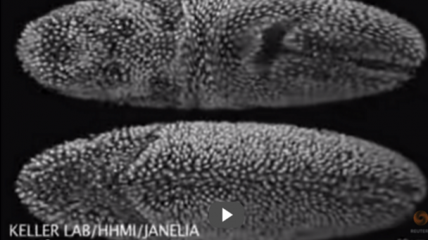
“The fruit fly has a very long and distinguished career in science. At a facility considered a Nirvana for scientists, researchers pursue greater understanding of biomedical processes, using test subjects like dragonflies and zebrafish.

PBS News Hour Science correspondent Miles O’Brien reports on how the Janelia Farm Research Campus supports groundbreaking basic research.”

Article on PBS website art

Published July 23, 2014 by PBS Newshour


Transcript

GWEN IFILL: Next: trying to better understand what’s happening in the brain of a fruit fly, a dragonfly, or a zebra fish, all part of a larger puzzle to learn more about how our own brains work.

NewsHour science correspondent Miles O’Brien has the first in our three-part series on the science of the brain.

MILES O’BRIEN: Oh, to be a fly on the wall at the Basic Research Facility scientist consider nirvana. You might see a Nobel Prize in the making or you might be subjected to this, the fruit fly version of a scary movie, the rapidly growing shadow of a predator homing in for the kill.

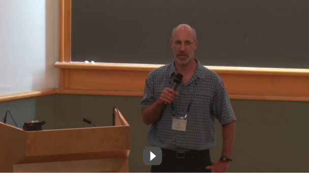
GWYNETH CARD, Howard Hughes Medical Institute-Janelia Farm Research Campus: My lab is really interested in how flies make decisions.

MILES O’BRIEN: Neuroscientist Gwyneth Card runs a laboratory at the Howard Hughes Medical ...

OnAir Post: What a fly’s brain tells us about our own minds

Unraveling embryonic development cell by cell

YouTube link

“New computer software to unravel embryonic development cell by cell”

Researchers at the Howard Hughes Medical Institute Janelia Research campus are using a new type of computer software to track and image how a nervous system develops in unprecedented detail. The new system is able to track individual cells during embryonic development, giving scientists a powerful to tool to create a blueprint of how brains form. Ben Gruber reports, Reuters.


Published Aug. 6, 2014 by Reuters


OnAir Post: Unraveling embryonic development cell by cell

Single molecules, cells, and super-resolution optics

https://www.youtube.com/watch?v=M2MOGnYe2lAVideo can’t be loaded because JavaScript is disabled: Single molecules, cells, and super-resolution optics (https://www.youtube.com/watch?v=M2MOGnYe2lA)

Nobel Laureate in Chemistry 2014: Eric Betzig, Janelia Research Campus, Howard Hughes Medical Institute, Ashburn, VA, USA.

From: The Nobel Lectures 2014, 2014-12-08.

Published on Jan. 8, 2015 by Vetenskapsakademien

OnAir Post: Single molecules, cells, and super-resolution optics

Imaging Life at High Spatiotemporal Resolution


https://www.youtube.com/watch?v=2R2ll9SRCeoVideo can’t be loaded because JavaScript is disabled: Eric Betzig: Imaging Life at High Spatiotemporal Resolution (https://www.youtube.com/watch?v=2R2ll9SRCeo)

In this lecture, held on 3/9/15 at UC Berkeley, Nobel Laureate Eric Betzig, describes three areas focused on addressing the challenges of high resolution imaging: super-resolution microscopy; plane illumination microscopy using non-diffracting beams; and adaptive optics to recover optimal images from within optically heterogeneous specimens.

Published on March 15, 2015 by UC Berkeley Events

OnAir Post: Imaging Life at High Spatiotemporal Resolution

Betzig and Hess: Developing PALM Microscopy


https://www.youtube.com/watch?v=GcQ24khZzvUVideo can’t be loaded because JavaScript is disabled: Eric Betzig and Harald Hess (Janelia Farm/HHMI): Developing PALM Microscopy (https://www.youtube.com/watch?v=GcQ24khZzvU)

During their 20-year friendship, Eric Betzig and Harald Hess, now at Janelia Farm/HHMI, worked together and separately, in academia and industry, before eventually joining forces to develop the first super-high-resolution PALM microscope.

They tell us the story of this journey and emphasize how their unusual and varied backgrounds provided the skills to complete the project.

Uploaded on Jan. 8, 2011 by iBioMagazine

OnAir Post: Betzig and Hess: Developing PALM Microscopy

Resolving Everything: Harald Hess

https://www.youtube.com/watch?v=esC6cr6qfs8Video can’t be loaded because JavaScript is disabled: Resolving Everything (https://www.youtube.com/watch?v=esC6cr6qfs8)

Harald Hess of the Janelia Research Campus of the Howard Hughes Medical Institute describes his path in basic physics, industry and biology and how the challenge of resolution in microscopy has guided and inspired his research.

Published April 1, 2015 by the National High Magnetic Field Laboratory

OnAir Post: Resolving Everything: Harald Hess

Inside the Lab: Karel Svoboda

https://www.youtube.com/watch?v=1jQIHrrbms8Video can’t be loaded because JavaScript is disabled: Inside the Lab: Karel Svoboda, Janelia Research Campus (https://www.youtube.com/watch?v=1jQIHrrbms8)

“An Exploration of Brain Dynamics”

Get a behind-the-scenes look at a day in the lab of neuroscientist Karel Svoboda as his group at the Howard Hughes Medical Institute’s Janelia Research Campus pushes to build a new type of microscope and uses a virtual reality system to learn how mice explore the world.

Published on July 28, 2014 by zincfinger23


OnAir Post: Inside the Lab: Karel Svoboda

Platform for large-scale neuroscience

https://www.youtube.com/watch?v=Gg_5fWllfgAVideo can’t be loaded because JavaScript is disabled: A platform for large-scale neuroscience – Jeremy Freeman (https://www.youtube.com/watch?v=Gg_5fWllfgA)

A platform for large-scale neuroscience” Jeremy Freeman (HHMI Janelia Farm Research Campus)

Filmed and recorded live at Spark Summit 2014 in San Francisco at the Westin St. Francis.

Published on July 17, 2014 by Apache Spark

OnAir Post: Platform for large-scale neuroscience

Tools for studying neurons: Loren Looger

https://www.youtube.com/watch?v=C7NUYgdzZ4YVideo can’t be loaded because JavaScript is disabled: CSHL Keynote Series; Dr. Loren Looger, HHMI Janelia Farm (https://www.youtube.com/watch?v=C7NUYgdzZ4Y)

“Tools for studying neurons, the only important cells in the brain” from the Glia in Health & Disease 7/19/2014

Presented by Dr. Loren Looger, HHMI Janelia Farm Part of Cold Springs Harbor Laboratory Keynote lecture series

Pulbished on July 21, 2014 by  CSHL Leading Strand

OnAir Post: Tools for studying neurons: Loren Looger

Paul Allen and Gary Marcus Fireside Chat: 2013 Annual Symposium


https://www.youtube.com/watch?v=ykBgh4nG88UVideo can’t be loaded because JavaScript is disabled: Paul Allen and Gary Marcus Fireside Chat: 2013 Annual Symposium (https://www.youtube.com/watch?v=ykBgh4nG88U)

Gary Marcus, a cognitive scientist at New York University, is the author of four books. He also writes a blog on artificial intelligence and science for The New Yorker. Paul G. Allen and his sister Jody Allen founded the Allen Institute for Brain Science.

A true visionary, Paul Allen was inspired by a host of leading scientists 10 years ago to move the needle on brain research.

Published on October 3, 2013  by Allen Institute for Brain Science

OnAir Post: Paul Allen and Gary Marcus Fireside Chat: 2013 Annual Symposium

Tutorials: Allen Institute Technical Tours

The twelve videos in this post are tutorials on the various Allen Brain Atlases published between 2011 and 2015.


Tutorial: Allen Human Brain Atlas

“This tutorial provides a brief orientation to the data and basic features of the Allen Human Brain Atlas, a multi-modal atlas of gene expression in the adult human brain.”

https://www.youtube.com/watch?v=x0PZ_SCXf-cVideo can’t be loaded because JavaScript is disabled: Webinar: Using the Allen Brain Atlas resources (October 2012) (https://www.youtube.com/watch?v=x0PZ_SCXf-c)


Allen Cell Types Database: Understanding the fundamental building blocks of the brain

“The Allen Institute for Brain Science is taking the first major scientific step to create a searchable standards database for the brain with the launch of the Allen Cell Types Database.”

https://www.youtube.com/watch?v=1GWyjxzxqIIVideo can’t be loaded because JavaScript is disabled: Allen Cell Types ...

OnAir Post: Tutorials: Allen Institute Technical Tours

Allen Data Base (video)

https://www.youtube.com/watch?v=1GWyjxzxqIIVideo can’t be loaded because JavaScript is disabled: Allen Cell Types Database: Understanding the fundamental building blocks of the brain (https://www.youtube.com/watch?v=1GWyjxzxqII)

“Allen Cell Types Database: Understanding the fundamental building blocks of the brain”

The Allen Institute for Brain Science is taking the first major scientific step to create a searchable standards database for the brain with the launch of the Allen Cell Types Database.

Published on May 14, 2015 by Allen Institute for Brain Science

OnAir Post: Allen Data Base (video)

Electron Microscopy Imaging

https://www.youtube.com/watch?v=WAkaLEM0gTQVideo can’t be loaded because JavaScript is disabled: Virtual Tour: Electron Microscopy Imaging (https://www.youtube.com/watch?v=WAkaLEM0gTQ)

A virtual journey through the Allen Institute for Brain Science’s Electron Microscopy (EM) imaging program

Published on April 9, 2014 by Allen Institute for Brain Science

OnAir Post: Electron Microscopy Imaging

Connectivity Dot-o-Gram

https://www.youtube.com/watch?v=3CvY-y2IPdgVideo can’t be loaded because JavaScript is disabled: Connectivity Dot-o-Gram (https://www.youtube.com/watch?v=3CvY-y2IPdg)

“This movie displays 21 mapping experiments from the Allen Mouse Brain Connectivity Atlas: a tool to investigate how different regions of the brain are connected. The density of axons at each voxel (dot) are displayed as overlapping circles color-coded by the area of the brain from which the axons are projecting. This animation shows how projections from different regions of the cortex divide the thalamus and striatum into distinct domains. The Allen Mouse Brain Connectivity Atlas is available to everyone online at www.brain-map.org.

Published on April 9, 2014 by Allen Institute for Brain Science

OnAir Post: Connectivity Dot-o-Gram

Cortico-thalamic connections

https://www.youtube.com/watch?v=6u0q5jIxR10Video can’t be loaded because JavaScript is disabled: Organization of the cortico-thalamic connections (https://www.youtube.com/watch?v=6u0q5jIxR10)

“This video shows a composite of 80 injection sites of a viral tracer in the cortex (round spheres) and their major projections into the thalamus. We can clearly see that the spatial pattern of the injections is conserved in the thalamus. This kind of information was only previously available in piecemeal form, but with the comprehensive, standardized data in the Allen Mouse Brain Connectivity Atlas, we can reproducibly measure and visualize this topography.”

Published on February 27, 2014 by Allen Institute for Brain Science


OnAir Post: Cortico-thalamic connections

Arthur Toga: 2013 Allen Symposium


https://www.youtube.com/watch?v=U8vesksXErsVideo can’t be loaded because JavaScript is disabled: Arthur Toga: 2013 Annual Symposium (https://www.youtube.com/watch?v=U8vesksXErs)

“Brain mapping and integrating multimodal data across subjects and projects.”

Dr. Arthur Toga is Provost Professor of Ophthalmology, Neurology, Psychiatry and the Behavioral Sciences, Radiology and Engineering at the University of Southern California, and serves as the inaugural Director of the USC Institute for Neuroimaging and Informatics. He also holds an appointment in the USC Viterbi School of Engineering. A leading authority on neuroimaging, informatics, mapping brain structure and function, and brain atlasing, Dr. Toga is the former Distinguished Professor of Neurology, University Professor, Vice Chair of the Department of Neurology, Associate Dean at the Geffen School of Medicine, and Co-Director of the UCLA Brain Mapping Center.

Published on October 3, 2013  by Allen Institute for Brain Science

OnAir Post: Arthur Toga: 2013 Allen Symposium

Cori Bargmann: 2012 Allen Symposium

https://www.youtube.com/watch?v=Blquxto1oIoVideo can’t be loaded because JavaScript is disabled: Cori Bargmann: 2012 Allen Institute for Brain Science Symposium (https://www.youtube.com/watch?v=Blquxto1oIo)

Published on November 12, 2012  by Allen Institute for Brain Science

Dr. Cori Bargmann, recent winner of the Kavli Prize in Neuroscience and a pioneer in methods of looking at C. elegans to uncover how neural circuits operate, presented the idea that particular classes of genes — neuropeptides to be specific — are good places to look for the genetic origin of behavior. Because much of the genome is conserved across species and throughout time, new behaviors may be created by redeploying old genes in different ways.

Uncovering the basic building blocks of behavior, she believes, is an unsolved question in neuroscience that is now becoming solvable.

“The question is not whether calbindin is expressed in the hippocampus, but whether it has something to do with the unique functions of human memory capabilities,” Bargmann said. “That is a hard question.” Certain kinds of molecules relate to innate behaviors that are shared across ...

OnAir Post: Cori Bargmann: 2012 Allen Symposium

Earl Miller: 2012 Allen Symposium

https://www.youtube.com/watch?v=6QjIKr_vIecVideo can’t be loaded because JavaScript is disabled: Earl Miller: 2012 Allen Institute for Brain Science Symposium (https://www.youtube.com/watch?v=6QjIKr_vIec)

Published on October 12, 2012  by Allen Institute for Brain Science

“Brain rhythms and cognition”

From physics to electrophysiology and imaging, Dr. Miller and his lab focus on broad and far-reaching approaches to neuroscience questions. This symposium talk focused on questions similarly posed by Sabine Kastner, namely the relationship between action potential timing and brain function. By recording neural activity in monkeys as they switch among two tasks, Miller found that over half of the recording sites in the network for one task also appear to participate in the network for the other task. This suggests that circuitry in the prefrontal cortex may overlap and that oscillations are the key to selecting appropriate networks for the task that needs to be performed. Miller showed that neural ensembles, or networks, in close proximity oscillate out of phase with one another to avoid being simultaneously activated. That is, the oscillations bind neural ensembles ...

OnAir Post: Earl Miller: 2012 Allen Symposium

Ed Lein: 2012 Allen Symposium

https://www.youtube.com/watch?v=pSqyRRec1cMVideo can’t be loaded because JavaScript is disabled: Ed Lein: 2012 Allen Institute for Brain Science Symposium (https://www.youtube.com/watch?v=pSqyRRec1cM)

Deciphering the mammalian brain transcriptome”

Published on November 12, 2012  by Allen Institute for Brain Science

The development, structure and function of our brains are guided by selective usage of the 20,000-odd genes in our genomes. Taking advantage of the Allen Institute’s anatomically and genomically comprehensive atlases of gene expression in the developing brain in species from mouse to human, Dr. Lein explores the molecular logic of gene expression in the brain. How do gene expression profiles relate to the functional and cellular architecture of the brain, and what makes the human brain unique? Focusing on the human neocortex, the outermost layer of the brain, Lein showed that molecular similarities reflect spatial proximity across the neocortex; such that neighboring cortical regions are more similar to one another than to more distant regions. Surprisingly, these molecular similarities vary across the cortex in a graded fashion, in contrast to models of cortical architecture ...

OnAir Post: Ed Lein: 2012 Allen Symposium

John Donoghue: 2012 Allen Symposium

“Neurotechnology: Fixing broken brains by decoding cortex”

Dr. Donoghue’s lab works in both basic and applied neuroscience, studying network computation on one end, and restoring lost motor cortical function on the other end. His work has culminated in BrainGate, a human brain-computer interface that senses neural activity in the motor cortex and decodes it to instruct a robotic arm or a cursor, reconnecting the cortex to lost action in the case of spinal cord injury or paralyzing disease such as amyotrophic lateral sclerosis. Donoghue showed video clips from his lab featuring long-paralyzed patients successfully navigating a robotic arm to drink their favorite beverage or guiding a computer cursor to communicate with loved ones. Among other noteworthy aspects of this work are the two notions that 1) the program actually decodes activity in the motor cortex rather than using a binary measure of activity (e.g., on or off) and 2) individual neurons in the motor cortex retain activity and function, even in quadriplegics who have not used this area of the brain in over a dozen years. While the functional utility of this work is self-evident, the value and applications to basic neuroscience are no less important. In going from a plan ...

OnAir Post: John Donoghue: 2012 Allen Symposium

Viviana Gradinaru, Ph.D., Next Generation Leader


https://www.youtube.com/watch?v=Awz4-Y_RKQQVideo can’t be loaded because JavaScript is disabled: Showcase 2014: Viviana Gradinaru, Ph.D., Next Generation Leader (https://www.youtube.com/watch?v=Awz4-Y_RKQQ)

Visualizing the activity and anatomy of brain circuits: optogenetic sensors and tissue clearing approaches

Published on September 26, 2014 by Allen Institute for Brain Science

OnAir Post: Viviana Gradinaru, Ph.D., Next Generation Leader

Science in 3D: 2015 Bioinformatics Festival

Day one videocast: Morning sessions included 3D printed Eyewire neurons by James Drake and Oculus fly through the brain.

Afternoon seesions included strategies for how to implement open source bio printing accessible and inexpensive… available for research. NIH 3D printer exchange is creating file formats for bioprinting cell types.


About the Festival

The NIAID Office of Cyber Infrastructure and Computational Biology (OCICB) is pleased to host our annual 2015 Bioinformatics & Computational Biosciences Festival, Science in 3D.

Despite the fast-growing applications for 3D technologies, they are not widely used in the biosciences at present. The festival will look at several aspects of 3D technologies including modeling, printing, and visualization. Speakers will explore bioprinting, physician and patient education, medical and surgical devices, augmented reality for medicine, and policies surrounding this exploding field. The festival will feature high-level presentations by subject matter experts from government agencies, academia, and the private sector.

Day one videocast   Day two videocast

Morning sessions included 3D printed Eyewire neurons by James Drake

and Oculus fly through the brain. See below from TED 2014 video.

https://www.youtube.com/watch?v=SU9etoLKgdIVideo can’t be loaded because JavaScript is disabled: Neurons in Space ...

OnAir Post: Science in 3D: 2015 Bioinformatics Festival

A look inside the brain in real time

The applications for real-time fMRIs start with chronic pain control and range into the realm of science fiction, but this technology is very real.

Filmed February 2008 at TED 2008
Uploaded to YouTube on March 27, 2008 by TED

TED Talks webpage

OnAir Post: A look inside the brain in real time

Development of the Human Brain

https://www.youtube.com/watch?v=-LiKX8BvBBIVideo can’t be loaded because JavaScript is disabled: Fueling Discovery: BrainSpan Atlas of the Developing Human Brain (https://www.youtube.com/watch?v=-LiKX8BvBBI)

Fueling Discovery: BrainSpan Atlas of the Developing Human Brain

“Ed Lein, an investigator at the Allen Institute for Brain Science, discusses how we are creating the BrainSpan Atlas of the Developing Human Brain and learning about how genes are used differently in the developing brain.”

Published on April 9, 2014 by Allen Institute for Brain Science


OnAir Post: Development of the Human Brain

What we’re learning from 5,000 brains

https://www.youtube.com/watch?v=ufUkAQOQaXUVideo can’t be loaded because JavaScript is disabled: Read Montague: What we’re learning from 5,000 brains (https://www.youtube.com/watch?v=ufUkAQOQaXU)

“Mice, bugs and hamsters are no longer the only way to study the brain. Functional MRI (fMRI) allows scientists to map brain activity in living, breathing, decision-making human beings. Read Montague gives an overview of how this technology is helping us understand the complicated ways in which we interact with each other.”

Filmed June 2012 at TED Global 2012 Uploaded to YouTube on September 24, 2012 by TED 

OnAir Post: What we’re learning from 5,000 brains

The quest to understand consciousness

https://www.youtube.com/watch?v=LMrzdk_YnYYVideo can’t be loaded because JavaScript is disabled: The quest to understand consciousness | Antonio Damasio (https://www.youtube.com/watch?v=LMrzdk_YnYY)

“Every morning we wake up and regain consciousness — that is a marvelous fact — but what exactly is it that we regain? Neuroscientist Antonio Damasio uses this simple question to give us a glimpse into how our brains create our sense of self.”

Filmed March 2011 at TED 2011 Uploaded to YouTube on December 18, 2011 by TED 

TED Talks webpage

OnAir Post: The quest to understand consciousness

How your brain tells you where you are

“How do you remember where you parked your car? How do you know if you’re moving in the right direction? Neuroscientist Neil Burgess studies the neural mechanisms that map the space around us, and how they link to memory and imagination.”

Filmed November 20111 at TEDSalon London 2011 Uploaded to YouTube on February 6, 2012 by TED 


TED Talks webpage

https://www.youtube.com/watch?v=Zd71719_G8YVideo can’t be loaded because JavaScript is disabled: Neil Burgess: How your brain tells you where you are (https://www.youtube.com/watch?v=Zd71719_G8Y)

“How do you remember where you parked your car? How do you know if you’re moving in the right direction? Neuroscientist Neil Burgess studies the neural mechanisms that map the space around us, and how they link to memory and imagination.”

Filmed November 20111 at TEDSalon London 2011 Uploaded to YouTube on February 6, 2012 by TED 

TED Talks webpage

OnAir Post: How your brain tells you where you are

What is so special about the human brain?

https://www.youtube.com/watch?v=_7_XH1CBzGwVideo can’t be loaded because JavaScript is disabled: What is so special about the human brain? | Suzana Herculano-Houzel (https://www.youtube.com/watch?v=_7_XH1CBzGw)

“The human brain is puzzling — it is curiously large given the size of our bodies, uses a tremendous amount of energy for its weight and has a bizarrely dense cerebral cortex. But: why? Neuroscientist Suzana Herculano-Houzel puts on her detective’s cap and leads us through this mystery. By making “brain soup,” she arrives at a startling conclusion.”

Filmed June 2013 at TEDGlobal 2013 Uploaded to YouTube on November 26, 2013 by TED 

TED Talks webpage

OnAir Post: What is so special about the human brain?

Could future devices read images from our brains?

“As an expert on cutting-edge digital displays, Mary Lou Jepsen studies how to show our most creative ideas on screens. And as a brain surgery patient herself, she is driven to know more about the neural activity that underlies invention, creativity, thought. She meshes these two passions in a rather mind-blowing talk on two cutting-edge brain studies that might point to a new frontier in understanding how (and what) we think.”

Filmed March 2013 at TED 2013 Uploaded to YouTube on March 3, 2013 by TED 

TED Talks webpage

https://www.youtube.com/watch?v=vNDhu2uqfdoVideo can’t be loaded because JavaScript is disabled: Mary Lou Jepsen: Could future devices read images from our brains? (https://www.youtube.com/watch?v=vNDhu2uqfdo)

OnAir Post: Could future devices read images from our brains?

Connectomics- Jeff Lichtman

https://www.youtube.com/watch?v=82tQ4ID-xNgVideo can’t be loaded because JavaScript is disabled: Jeff Lichtman: Connectomics: Mapping the Brain | Harvard Department of Physics (https://www.youtube.com/watch?v=82tQ4ID-xNg)

Despite intense interest in the ways brains work, we still have quite a rudimentary understanding of this organ, especially compared to our knowledge of the other organ systems in the body.

One central problem is that brain function is based on a much more complicated cellular organization than found in any other part of the body.

Published July 13, 2013 by Harvard University

Synopsis

Despite intense interest in the ways brains work, we still have quite a rudimentary understanding of this organ, especially compared to our knowledge of the other organ systems in the body. One central problem is that brain function is based on a much more complicated cellular organization than found in any other part of the body. The brain contains billions of nerve cells and these are interconnected by trillions of synapses in a vast wiring diagram. This wiring diagram has ...

OnAir Post: Connectomics- Jeff Lichtman

3 clues to understanding your brain


https://www.youtube.com/watch?v=Rl2LwnaUA-kVideo can’t be loaded because JavaScript is disabled: 3 clues to understanding your brain | VS Ramachandran (https://www.youtube.com/watch?v=Rl2LwnaUA-k)

“Vilayanur Ramachandran tells us what brain damage can reveal about the connection between celebral tissue and the mind, using three startling delusions as examples.”

Filmed march 2007 at TED 2007 Uploaded to YouTube on October 23, 2007 by TED  

TED Talks webpage

OnAir Post: 3 clues to understanding your brain

Understanding the Mind by Mapping the Brain


https://www.youtube.com/watch?v=igDuigbb-9I

“Understanding the Mind by Mapping the Brain”

Jacopo Annese, Director of the Brain Observatory at the University of California, San Diego is on a quest to collect, dissect, and digitize images of the human brain for the Digital Brain Library, which was launched with support from the National Science Foundation. Annese and his team look for connections, mapping brain structure and connecting it to human behavior.

NSF BRAIN Initiative Science Nation – August 4, 2011

Description

Neuroanatomist Jacopo Annese is looking for 1,000 brains. The Director of the Brain Observatory at the University of California, San Diego is on a quest to collect, dissect, and digitize images of the human brain for the Digital Brain Library, which was launched with support from the National Science Foundation. Annese and his team look for connections, mapping brain structure and connecting it to human behavior. He believes that with a large enough catalog of brains preserved as virtual models, scientists can explore the organ in ways unheard of, revealing new insights into what makes the brain tick. Annese is collecting data on the lives of the people who have already donated brains. He is also studying the behavior of people ...

OnAir Post: Understanding the Mind by Mapping the Brain

Mapping the mouse brain in 3-D

https://www.youtube.com/watch?v=kbv9qoJLheQVideo can’t be loaded because JavaScript is disabled: Mapping the mouse brain in 3-D (https://www.youtube.com/watch?v=kbv9qoJLheQ)

The mapping of the mouse brain is giving researchers like Partha Mitra a better understanding of how neurons are connected and how they communicate across the regions of the brain.


NSF BRAIN Initiative Published APRIL 2, 2014

OnAir Post: Mapping the mouse brain in 3-D

Picturing the Brain – Paul Thompson

https://www.youtube.com/watch?v=qkYEfWazIfcVideo can’t be loaded because JavaScript is disabled: Picturing the Brain (https://www.youtube.com/watch?v=qkYEfWazIfc)

Dr. Paul Thompson talks about the work that he does at the Lab of Neuro Imaging (LONI) at UCLA.

He discusses brain health, the latest brain imaging technology and projects like ENIGMA, which involves a world-wide effort to create a brain database.

Published on July 13m 2013 by NIBIBgov

OnAir Post: Picturing the Brain – Paul Thompson

Skip to toolbar