Neural Recording Overview

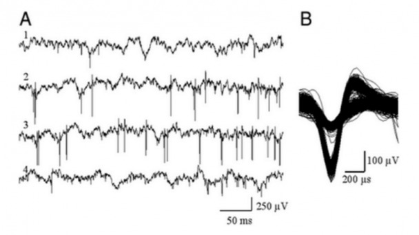
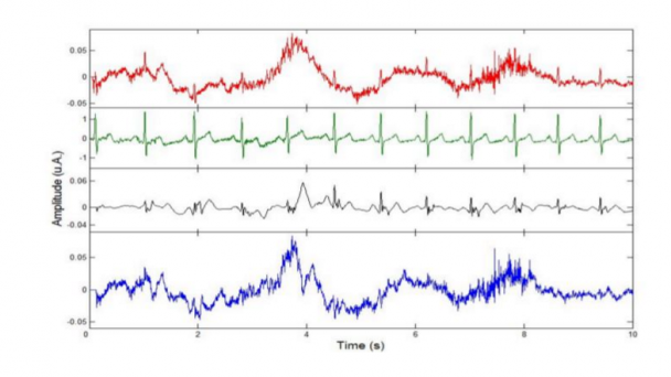
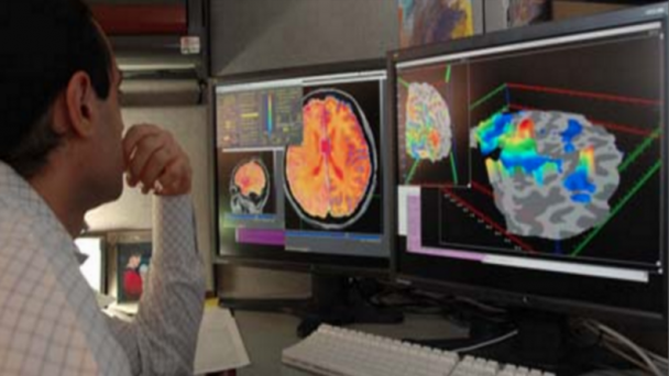
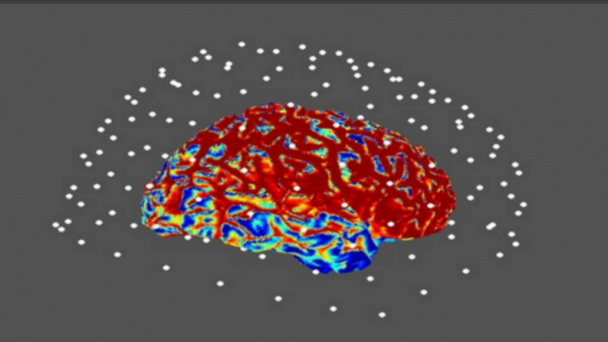
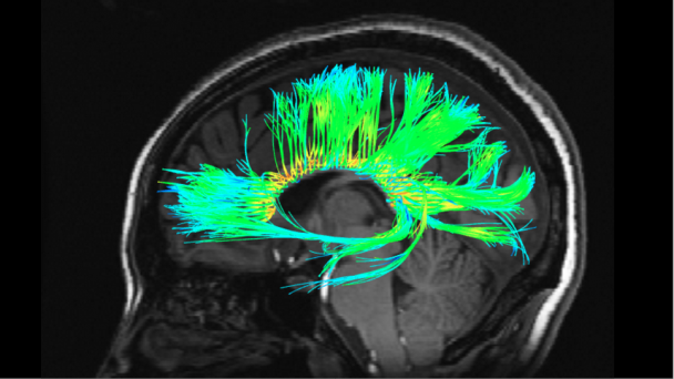
Common neuroimaging methods for measuring brain function include: Positron emission tomography (PET), Functional magnetic resonance imaging (fMRI), multichannel electroencephalography (EEG), magnetoencephalography (MEG), near infrared spectroscopic imaging (NIRSI), and Single-photon emission computed tomography (SPECT). Single neuron measurement normally uses a microelectrode system.

OnAir Post: Neural Recording Overview

Single-unit and multi-electrode recording

When a neuron generates an action potential, the signal propagates down the neuron as a current which flows in and out of the cell through excitable membrane regions in thesoma and axon. A microelectrode is inserted into the brain, where it can record the rate of change in voltage with respect to time.

OnAir Post: Single-unit and multi-electrode recording

Electroencephalography

EEG measures voltage fluctuations resulting from ionic current within the neurons of the brain. In clinical contexts, EEG refers to the recording of the brain's spontaneous electrical activity over a period of time, as recorded from multiple electrodes placed on the scalp. Diagnostic applications generally focus on the spectral content of EEG, that is, the type of neural oscillations that can be observed in EEG signals.

OnAir Post: Electroencephalography

Functional magnetic resonance imaging

This technique relies on the fact that cerebral blood flow and neuronal activation are coupled. When an area of the brain is in use, blood flow to that region also increases.

Also see Scholarpedia. Curator Karl Friston (2007)

From Wikipedia Oct. 16, 2015

OnAir Post: Functional magnetic resonance imaging

Magnetoencephalography

Typically MEG sensors are housed in a helmet shaped container (or dewar) in which the subject places their head during the measurement process.

OnAir Post: Magnetoencephalography

Skip to toolbar