Decoding and Modulation of Human Language

Principal Investigators: Behnaam Aazhang, PhD – Rice and Nitin Tandon, MD – UT Health Title: Micro-scale Real-time Decoding and Closed-loop Modulation of Human Language BRAIN Category: Neuroengineering and Brain-inspired concepts and design

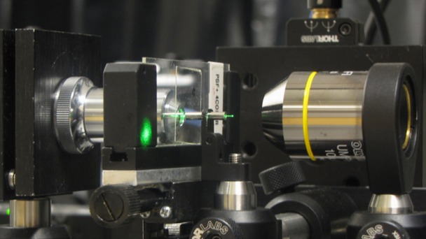
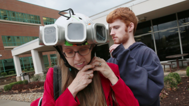
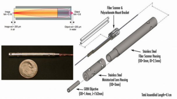
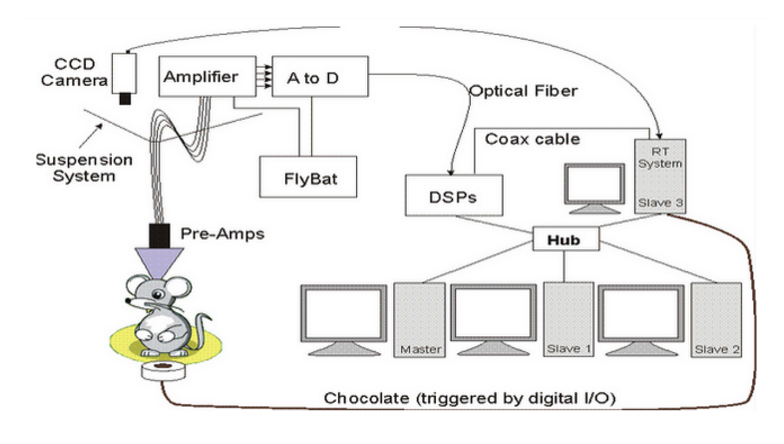
The engineering objective is to develop biocompatible microchips to vastly enhance our insight into language and other cognitive processes and learning. Miniaturized microchips in silicon technology will be developed that can record neural signals, digitize them, and transmit the signals to an in vitro receiver wirelessly.

Abstract

Award Number: #1533688

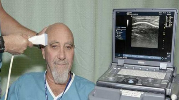
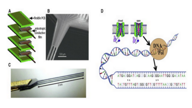
Humans produce language, which is a defining characteristic of our species and our civilization. We can select words precisely out of a large lexicon with remarkably low error rates. It is perhaps not surprising that this complex speech production system is easily affected by disease. Brain damage induced language disorders affect millions of Americans, and there is little hope of remediation. Research on the anatomical, physiological, and computational bases of speech production has made important strides in recent years but this has been limited by a glaring lack of information on the dynamics of the process. This limitation results from the low spatio-temporal resolution of the available tools to collect data and the effectiveness of the current tools for analysis. Our driving vision ...

OnAir Post: Decoding and Modulation of Human Language

Imaging synaptic activity using super-resolution cannula microscopy

Principal Investigator: Rajesh Menon – Utah Neuroscience Title:Imaging synaptic activity deep in the brain using super-resolution cannula microscopy” BRAIN Category: Neuroengineering and Brain-inspired concepts and design (#1532591)

Objective: This project will develop a tool for high-resolution (<100-nm) imaging of synapses in freely moving animals for neuronal studies. It will accomplish this goal by the development and integration of compact and lightweight cannula microscopy with in vitro fluorescence imaging with accompanying technology and methodologies for imaging synapses.

Abstract

Award Number#1533611

Objective: This project will develop a tool for high-resolution (<100-nm) imaging of synapses in freely moving animals for neuronal studies. It will accomplish this goal by the development and integration of compact and lightweight cannula microscopy with in vitro fluorescence imaging with accompanying technology and methodologies for imaging synapses.

Non-Technical

The long-term vision of this project is to image with high resolution deep inside the brain of freely moving mice using inexpensive technologies so as to elucidate the fundamental basis of information processing and memory. Changes in synaptic strength at specific synapses are thought to underlie memory encoding and storage, yet there is very little experimental evidence for this theory in the intact brain due to technical limitations of visualizing the specific synaptic pattern involved in experience-dependent ...

OnAir Post: Imaging synaptic activity using super-resolution cannula microscopy

Magnetic Particle Imaging (MPI)

Principal Investigator: Lawrence Wald Neuroscience@Harvard Title: “Magnetic Particle Imaging (MPI) for Functional Brain Imaging in Humans” BRAIN Category: Next Generation Human Imaging (RFA MH-14-217)

The Wald team plans to use an iron-oxide contrast agent to track blood volume, which will permit dramatically more sensitive imaging of human brain activity than existing methods.

NIH Webpages

Schematic set up and operating principle of the Magnetic Particle Imaging technology. Phillips MPI.

Project Description

In this planning grant we propose several engineering developments to advance Magnetic Particle Imaging (MPI) to replace MRI as the next-generation functional brain imaging tool for human neuroscience. We assemble a group of technology experts to solve a myriad of identified and unidentified barriers, we employ simulation and bench-top experiments to characterize and test solutions for these technical obstacles and validate solutions by bench testing specific sub-sections of the imager. Finally we simulate the overall performance of the planned device and assess its benefit for human functional brain imaging. MPI is a young but extremely promising technology that uses the nonlinear magnetic response of iron- oxide nanoparticles to localize their presence in the body. MPI directly detects the nanoparticle’s magnetization rather than using secondary effects on the Magnetic Resonance relaxation times. ...

OnAir Post: Magnetic Particle Imaging (MPI)

Imaging Brain Function with Portable MRI

Principal Investigator: Michael Garwood Institute for Translational Neuroscience, University of Minnesta Title: “Imaging Brain Function in Real World Environments & Populations with Portable MRI” BRAIN Category: Next Generation Human Imaging (RFA MH-14-217)

By employing smaller, less cumbersome magnets than used in existing MRI, Dr. Garwood and colleagues will create a downsized, portable, less expensive brain scanner.

NIH Webpages

Project Description

Functional magnetic resonance imaging (fMRI) continues to play a critical role in understanding the human brain. Yet current fMRI technology is far less than ideal for studying brain function due to the unnatural environment and restricting space of the magnet bore. Furthermore, fMRI cannot be performed on subjects who have metallic implants in their body (e.g., the elderly, soldiers and veterans), or who are impaired by certain physical disabilities as occurs in a variety of neurological and vestibular disorders. Finally, due to its expense and infrastructure requirements, MRI’s predominant accessibility to wealthier institutions has resulted in a highly biased subject sampling and a shortage of studies in non-western environments and cultures. The general methodology used to obtain MR images today is essentially the same as that used approximately 4 decades ago. One major drawback of such methodology is that the tolerated magnetic field variation over the brain is ...

OnAir Post: Imaging Brain Function with Portable MRI

Advancing MRI & MRS Technologies

Principal Investigator: Wei Chen Institute for Translational Neuroscience, University of Minnesota Title: “Advancing MRI & MRS Technologies for Studying Human Brain Function and Energetics” BRAIN Category: Next Generation Human Imaging (RFA MH-14-217)

Dr. Chen’s team will achieve unprecedented higher resolution magnetic resonance imaging and spectroscopy scanning by integrating ultra-high dielectric constant material and ultra-high-field techniques.

NIH Webpages

Engineering for Ultrahigh field Magnetic Resonance Imaging (MRI) and Spectroscopy (MRS)

Project Description

Magnetic resonance (MR) imaging (MRI) and in vivo MR spectroscopy (MRS) techniques have become indispensable tools for imaging brain structure, function, connectivity, neurochemistry and neuroenergetics, and for investigating neurological disorders. However, it remains a challenge to achieve superior MRI/MRS detection sensitivity, spatial and temporal imaging resolutions adequate for addressing fundamental and challenging neuroscience questions even with the most advanced technology. The prevailing paradigms for improving MRI/MRS performance largely invoke increasing the magnetic field strength, which may have reached practically achievable limits for human studies due to many technological and safety (i.e., high specific absorption rate (SAR)) concerns, and increasing the receiver channel count which is also ultimately limited due to noise characteristics of coils of decreasing size. To alleviate these major limitations, this R24 proposal relies on the interdisciplinary research efforts and ...

OnAir Post: Advancing MRI & MRS Technologies

MRI Neuro-Electro-Magnetic Oscillations

Principal Investigator: Allen W Song Duke Institute for Brain Sciences Title: “Path Toward MRI with Direct Sensitivity to Neuro-Electro-Magnetic Oscillations” BRAIN Category: Next Generation Human Imaging (RFA MH-14-217)

Dr. Song’s group will develop a scanner technology sensitive enough to image brain activity in high resolution by directly tuning in the electromagnetic signals broadcast by neurons.

NIH Webpages

Spiral imaging is a fast MRI technique that is widely used in functional MRI. It is, however, vulnerable to spatial and temporal variations of the static magnetic field (B0) caused by susceptibility effects, subject motion, physiological noise, and system instabilities, resulting in blurring artifacts. To address these issues, we have developed a novel off-resonance correction method, based on k-space energy spectrum analysis (KESA), for inherent and dynamic B0 mapping and deblurring in spiral imaging. This method can generate a B0 map from the k-space data at each time point, without requiring any additional data acquisition or pulse sequence modification, and correct for the blurring caused by both spatial and temporal B0 variations, resulting in a high spatial and temporal fidelity.

Project Description

In response to the NIH RFA-MH-14-217 on “Planning for Next Generation Human Brain Imaging”, we propose a comprehensive plan to organize the ...

OnAir Post: MRI Neuro-Electro-Magnetic Oscillations

Vascular Interfaces for Brain Imaging

PI: Robert Desimone Massachusetts Institute of Technology Title: “Vascular Interfaces for Brain Imaging and Stimulation” BRAIN category: Next Generation Human Imaging (RFA MH-14-217)

Dr. Desimone’s project will access the brain through its network of blood vessels to less invasively image, stimulate and monitor electrical and molecular activity than existing methods.

NIH Webpages

Sketch showing constitution of blood vessels inside the brain. Credit: Armin Kübelbeck

Project Description

Functional MRI (fMRI), EEG, and other completely noninvasive modalities for large-scale imaging of human brain activity have pioneeringly revealed many human brain functions, but cannot reach the single-neuron, single-spike level of neural code analysis possible in animals obtained using electrodes. This is partly due to the indirect methods of observation employed (e.g., blood flow for fMRI) and due to blurring of signals over distance by the skull (e.g., for EEG). In contrast, invasive approaches such as trans-cranially implanted multi- electrode arrays can achieve single-cell, single-spike resolution, but they necessitate opening of the skull – and, for implanted arrays, damage of the brain tissue – limiting utility to a small fraction of the population, those undergoing neurosurgery for some intractable brain disorder that justifies the risk. Trans-cranially implanted arrays also degrade i performance over time ...

OnAir Post: Vascular Interfaces for Brain Imaging

Ultrasonic neuromodulation in vivo

PI: Doris Ying  Tsao California Institute of Technology Title: “Dissecting human brain circuits in vivo using ultrasonic neuromodulation” BRAIN category: Next Generation Human Imaging (RFA MH-14-217)

In rodents, monkeys and eventually humans, Dr. Tsao’s team will explore use of non-invasive, high resolution ultrasound to impact neural activity deep in the brain and modify behavior.

NIH Webpages


Project Description

A dream of neuroscience is to be able to non-invasively modulate any given region of the human brain with high spatial resolution. This would open new horizons for understanding human brain function and connectivity, and create completely new options for the non-invasive treatment of brain diseases such as intractable epilepsy, depression, and Parkinson’s disease. Current non-invasive brain stimulation methods such as transcranial magnetic stimulation (TMS) and transcranial electrical stimulation (TES) can be applied only to superficial cortical areas, with crude 1 cm-scale resolution, limits placed upon these techniques by fundamental physics. Ultrasonic neuromodulation, the use of ultrasound as an energy modality to affect the activity of the brain, could overcome these limitations and thereby transform both basic and clinical human neuroscience. In fact, the engineering challenge of non-invasively focusing ultrasound to mm-sized regions, either shallow or deep in the brain, has been solved: clinical studies have already demonstrated ...

OnAir Post: Ultrasonic neuromodulation in vivo

Micro-Dose, Wearable PET Brain Imager

Principal Investigator:  Julie Brefczynski-Lewis WVU Center for Neuroscience Title: Imaging the Brain in Motion: The Ambulatory Micro-Dose, Wearable PET Brain Imager BRAIN Category: Next Generation Human Imaging (RFA MH-14-217)

Dr. Brefczynski-Lewis and co-workers will engineer a wearable PET scanner that images activity of the human brain in motion – for example, while taking a walk in the park.

NIH Webpages

Brefczynski-Lewis, assisted by graduate student Chris Bauer, dons her PET-helmet prototype to demonstrate the device’s portability. (Lois Raimondo/For The Washington Post)

Project Description

Our vision is to design the first truly mobile molecular brain imager that can be used on healthy subjects to study the functioning of the human brain during motion. The ultimate goal is to be able to image subjects during a proverbial “walk in the park” and other natural activities. We selected PET technology as the most likely to succeed in the next decade to provide the desired functionality of such a brain imager. While MRI is an exceptionally powerful and versatile imaging modality, and there are even upright MRIs for structural brain scans, for functional fMRI scans the subjects must stay still and in horizontal position inside a narrow bore of a strong-field MRI magnet. What we ...

OnAir Post: Micro-Dose, Wearable PET Brain Imager

Multiplexed Nanoscale In Situ Proteomics

Edwards S. Boyden Massachusetts Institute of Technology Title: “Ultra-Multiplexed Nanoscale In Situ Proteomics for Understanding Synapse Types” BRAIN category: Tools for Cells and Circuits (RFA MH-14-216)

Dr. Boyden’s team will simultaneously image both the identities and locations of multiple proteins within individual synapses – made possible by a new technique called DNA-PAINT.

NIH Webpages

DNA-PAINT super-resolution image of microtubules inside a fixed HeLa cell using Atto 655–labeled imager strands (10,000 frames, 10-Hz frame rate). Inset, labeling and imaging schematic for DNA-PAINT in a cellular environment. From Neuron doi:10.1038/nmeth.2835

Project Description

Significant work is ongoing to reveal how different cell types in the brain contribute to behavior and pathology, and how they change in plasticity and disease, empowered by new genetic, optical, and physiological tools. However, the functional activity and dysregulation of neuronal circuits relies critically on the in situ molecular composition of neuronal synapses. Although it is clear that the properties of a given synapse are determined by, amongst other things, the specific types of cells that are thus connected, far less is known about the diversity of synapse types in the brain than cell types, perhaps because this is an intrinsically proteomic problem: a given neuron might make many ...

OnAir Post: Multiplexed Nanoscale In Situ Proteomics

Nontoxic transsynaptic tracing

Principal Investigator: Ian Wickersham MIT Neuroscience Title: “Novel technologies for nontoxic transsynaptic tracing” BRAIN Category: Tools for Cells and Circuits (RFA MH-14-216)

Dr. Wickersham and colleagues will develop nontoxic viral tracers to assist in the study of neural circuitry underlying complex behaviors.

NIH Webpages

(a–c) Injection site in mouse somatosensory thalamus of an equal mixture of two deletion-mutant rabies viral vectors: VSVG-enveloped vector encoding mOrange2 (yellow), and an RVG-enveloped vector encoding mCherry (red). The VSVG-enveloped vector prolifically infects thalamic neurons at the injection site, whereas the RVG-enveloped one infects far fewer cells locally. (d–f) In the somatosensory cortex of the same mouse, mOrange2-filled thalamocortical axons ascend to layer 4 and densely ramify among the apical dendrites of mCherry-filled layer 6 corticothalamic cells retrogradely infected by the co-injected retrograde virus. Scale bar, 200 μm, applies to all panels.

Project Description

Genetic tools have dramatically increased the power and resolution of neuroscientific experiments, allowing monitoring and perturbation of specific neuronal populations within the brain, often in the context of complex cognitive and behavioral paradigms. However, the usefulness of these tools is limited by the available means of delivering them in circuit-specific ways, a major drawback in view of the critical importance of specific connectivity ...

OnAir Post: Nontoxic transsynaptic tracing

Activity measurement at single cell

Principal Investigator: Craig Forest Georgia Institute of Technology Title: “In-vivo circuit activity measurement at single cell, sub-threshold resolution” BRAIN Category: Tools for Cells and Circuits (RFA MH-14-216)

Dr. Forest’s team will use a newly developed robot guided technique to measure precise changes in electrical activity from individual neurons that are connected over long distances across the brain, to understand how these connections change when our brains go into different states, such as sleeping and waking.

NIH Webpages


Whole-cell patch clamp electrophysiology of neurons, although a gold standard technique for high-fidelity analysis of the biophysical mechanisms of neural computation and pathology, requires great skill to perform. We have developed a simple robot that automatically performs patch clamping in vivo, algorithmically detecting cells by analyzing the temporal sequence of electrode impedance changes. We demonstrate good yield, throughput, and quality of recording in mouse cortex and hippocampus..

Project Description

Neurons communicate information through fluctuations in the electrical potentials across their cellular membranes. Whole-cell patch clamping, the gold standard technique for measuring these fluctuations, is something of an art form, requiring great skill to perform on only a few cells per day. Thus, it has been primarily limited to in vitro experiments, a few in ...

OnAir Post: Activity measurement at single cell

Calcium sensors for molecular fMRI

PI: Alan Jasanoff Massachusetts Institute of Technology Title: “Calcium sensors for molecular fMRI” BRAIN category: Large-Scale Recording-Modulation – New Technologies (RFA NS-14-007)

Dr. Jasanoff’s team will synthesize calcium-sensing contrast agents that will allow functional magnetic resonance imaging (fMRI) scans to reveal activity of individual brain cells.

NIH Webpages

Project Description

The development of minimally invasive direct readouts of neural activity is one of the greatest challenges facing neuroscience today. Our recent work has shown that it is possible to perform high resolution functional magnetic resonance imaging (fMRI) of molecular-level phenomena using MRI contrast agents sensitive to hallmarks of neurotransmitter release. An even more valuable contribution would be the creation of calcium sensors suitable for molecular fMRI of intracellular neural signaling processes. Functional imaging performed with these sensors would combine the noninvasiveness and whole-brain coverage of MRI with the molecular specificity and broad applicability of established optical calcium neuroimaging techniques. Calcium-dependent fMRI will be a breakthrough technique for analysis of neural circuits in animals, with potential longer term applications in humans. The technique could achieve cellular resolution in conjunction with ultrahigh field MRI scanners and cell labeling techniques. A major hurdle in realizing this advance is the creation of effective calcium-dependent MRI contrast agents, however. This proposal describes ...

OnAir Post: Calcium sensors for molecular fMRI

Next generation imaging in vivo

Principal Investigator: Elly  Nedivi Massachusetts Institute of Technology Title: “Next generation high-throughput random access imaging, in vivo” BRAIN category: Large-Scale Recording-Modulation – Optimization (RFA NS-14-008)

Dr. Nedivi’s team proposes a new imaging technology to simultaneously record activity at each of the thousands of synapses, or communication points, on a single neuron.

NIH Webpages

https://www.youtube.com/watch?v=lHLSFhp5HawVideo can’t be loaded because JavaScript is disabled: Neurotech 1: Multi-Photon Microscopy (https://www.youtube.com/watch?v=lHLSFhp5Haw)

Project Description

The goal of this proposal is to develop new methods for high speed monitoring of sensory-driven synaptic activity across all inputs to single living neurons in the context of the intact cerebral cortex. Although our focus is on understanding how synaptic inputs are integrated across a single neuron embedded in an intact circuit, the next generation random access imaging technology we propose is more broadly applicable for monitoring multi-cellular activity representing large intra-and inter areal neuronal networks. The approach improves on the speed and sensitivity of current random-access technology by nearly 2 orders of magnitude, enabling high- throughput interrogation of up to 104 independent locations within a fraction of a millisecond and compatible with imaging using next generation voltage sensitive indicators. ...

OnAir Post: Next generation imaging in vivo

Rapid imaging of a living brain

Using a new high-speed, high-resolution imaging method, researchers at Washington University were able to see blood flow, blood oxygenation, oxygen metabolism, and other functions inside a living mouse brain at faster rates than ever before.

The new method is called “photoacoustic microscopy” (PAM), a single-wavelength, pulse-width-based technique developed by Lihong Wang, PhD, the Gene K. Beare Professor of Biomedical Engineering in the School of Engineering & Applied Science, and his team.

Schematic of fast functional photoacoustic microscopy (PAM) of the mouse brain. OAC, optical-acoustic combiner; PBS, polarizing beam splitter; UT, ultrasonic transducer. (credit: Junjie Yao et al./Nature).

Press Release

High-tech method allows rapid imaging of functions in living brain

Washington University in St. Louis 3/30/15 by Beth Miller

These images show fast functional photoacoustic microscopy of the mouse brain. Figure (d) shows a representative x-y projected brain vasculature image through an intact skull. Figure (e) shows a representative enhanced x-z projected brain vasculature image. Figure (f) shows photoacoustic microscopy of oxygen saturation of hemoglobin in the mouse brain, acquired by using the single-wavelength pulse-width-based method with two lasers.

​Researchers studying cancer and other invasive diseases rely on high-resolution imaging to see tumors and ...

OnAir Post: Rapid imaging of a living brain

Circuitry Underlying Memory replay

Principal Investigator: Ivan Soltesz UC Irvine Neuroscience Title: “Towards a Complete Description of the Circuitry Underlying Memory replay” BRAIN Category: Understanding Neural Circuits (RFA NS-14-009)

Dr. Soltesz’s team will combine computer brain modeling and large-scale recordings of hundreds of neurons to understand how the brain generates sharp-wave-ripples, a neuronal activity pattern essential for learning and memory.

NIH Webpages

How GABAergic interneurons in the hippocampus choose their postsynaptic partners

Project Description

The function of a brain region is an emergent property of many cell types. The criteria needed to understand a network have been established in studies of invertebrate “simple” networks, but there has not yet been an attempt to provide such a full, mechanistic understanding of any network in the vertebrate brain. We believe that the time is now ripe for such an effort. Specifically, we propose to understand how the CA3 network in the hippocampus generates sharp-wave-ripples (SWR). These events are of great interest because of their cognitive function: they represent replay of episodic memory sequences and are required for subsequent memory recall, as demonstrated at the behavioral level. Our efforts to understand the SWR will build on previous work establishing the cell types of the hippocampus. However, to meet ...

OnAir Post: Circuitry Underlying Memory replay

Optimization of 3-photon microscopy

Principal Investigator: Chris Xu Cornell University Title: “Optimization of 3-photon microscopy for Large Scale Recording in Mouse Brain” BRAIN Category: Large-Scale Recording-Modulation – Optimization (RFA NS-14-008)

Dr. Xu and his collaborators will build new lasers and lenses to use three-photon microscopy to watch neuronal activity far deeper inside the brain than currently possible.

NIH Webpages

Project Description

The goal of this research program is to optimize three-photon fluorescence microscopy (3PM) for large scale, noninvasive, volumetric imaging of neuronal activity. To leverage the superb performance of green-fluorescent protein based genetically engineered Ca-probes (e.g., GCaMPs), 3PM at the 1300-nm spectral window will be developed, which not only preserves the tissue penetration capability of 3PM at the longer excitation wavelength but also enables a wide variety of blue and green fluorophores, including a number of fluorescent proteins and Ca-indicators, to be excitable via three-photon excitation. To improve the signal-to-noise ratio (SNR) so that a practical frame rate can be achieved for imaging dynamic brain activity even at a penetration depth of 1.1 mm or beyond, new objective lenses will be designed and fabricated that will collect the signal efficiently at depth. In additin, the lens design will also support convenient integration with adaptive optics (AO), with the goal of making ...

OnAir Post: Optimization of 3-photon microscopy

Protein-based Voltage Probes

Principal Investigator: Vincent Allen Pieribone Yale Neuroscience Title: “Development of Protein-based Voltage Probes” BRAIN Category: Large-Scale Recording-Modulation – Optimization (RFA NS-14-008)

Dr. Pieribone and his team will optimize fluorescent voltage probe technology, to allow scientists to measure the activity of thousands of neurons using only a camera and a microscope.

NIH Webpages

Fluorogenetic Voltage Sensors. From website.

Project Description

The use of genetically encoded fluorescent activity probes represent the most advanced method to monitor the electrical activity of networks of neurons without using electrodes. While genetically encoded calcium indicators have been evolved to produce robust signals in a variety of different neuronal preparations, fluorescent probes of membrane potential have not been well evolved. Current voltage probes, while finally in expanded use, will need considerable improvement if the goal of recording the activity of a large number of neurons simultaneously in vivo is to be achieved. The goal of this project is to discover protein-based fluorescent voltage probes with signal to noise characteristics that allow routine optical recording of action potentials from single cortical neurons in vivo. We are seeking probes with significantly improved signal to noise characteristics, red-shifted fluorescence spectra, faster on and off rates and better plasma membrane expression. This ...

OnAir Post: Protein-based Voltage Probes

Protein voltage sensor imaging in vivo

Principal Investigator: Mark J Schnitzer Stanford Neuroscience Title: “Protein voltage sensors: kilohertz imaging of neural dynamics in behaving animals” BRAIN Category: Large-Scale Recording-Modulation – Optimization (RFA NS-14-008)

Dr. Schnitzer and his team have created a new system for developing optical voltage sensors, which will allow scientists to simultaneously record firing of large groups of neurons or electrical activity in precise locations inside of neurons, such as synapses.

NIH Webpages

The diagrams show a hypothetical protein (orange oval) and the formation of an active site, which is due to a voltage-induced conformational change that is mediated by the defined regions of the protein (green and yellow cylinders). a | Charged amino acids may move within membranes in response to changes in voltage. The side groups of Asp and Arg are shown. b | Reorientation of an intrinsic residue dipole, such as Tyr, through changes in the field. c | An alpha-helix that is the length of the membrane (red to blue gradient) has a dipole moment that is equivalent to the length of the helix that separates plusminus0.5 electronic charges (e0); therefore, it can also reorientate when the field is changed. The oval that is attached to the alpha-helix ...

OnAir Post: Protein voltage sensor imaging in vivo

Multi-area two-photon microscopy

Principal Investigator: Fritjof Helmchen Zurich Brain Research Institute Title: “Multi-area two-photon microscopy for revealing long-distance communication between multiple local brain circuits” BRAIN Category: Large-Scale Recording-Modulation – Optimization (RFA NS-14-008)

Dr. Helmchen and his colleagues propose a system to simultaneously record neuronal activity in four different areas of the neocortex and discover how brain cells in different regions interact during specific behaviors.

NIH Webpages

Two-photon laser-scanning microscopy has become the method of choice for imaging nerual dynamics on the cellular level in the intact brain. The particular advantage of 2-photon microscopy is its reduced sensitivity to light scattering so that individual cells with their dendrites can be resolved several hundreds of micrometers deep in neural tissue in living animals.

Project Description

Two-photon microscopy is a widely used, key method for functional imaging of cellular activity in living animals. Most recently, in vivo calciu imaging experiments have started to reveal the spatiotemporal activity patterns that occur in various areas of the neocortex during head-fixed mouse behavior. Typically, however, the field-of- view for imaging cellular activity is fairly small, on the order of a few hundred micrometers. This restriction limits the size of neuronal networks that can be studied and thus leaves open ...

OnAir Post: Multi-area two-photon microscopy

Electrophysiological Recording and Control

Principal Investigator: Baldwin Goodell Graymatter Research Title: “Large-Scale Electrophysiological Recording and Optogenetic Control System” BRAIN Category: Large-Scale Recording-Modulation – Optimization (RFA NS-14-008)

Dr. Goodell and his colleagues aim to develop optrodes, which are implantable columns of lights and wires for simultaneous electrical recording of neurons and delivery of light flashes to multiple brain areas.

NIH Webpages

Example data collected from 4 electrodes during a single recording session. A: short epoch of raw data sampled from 4 electrodes in area 7a of the posterior parietal cortex of an alert monkey. The signals are broadband (1 Hz to 10 kHz). Extracellular action potentials are visible as negative going spikes in the signals. B: plot of 1,000 superimposed waveforms extracted from the high-pass filtered signal on channel 3. The high-amplitude waveforms reveal a single unit that is well separated from the lower amplitude multiunit activity.

Project Description

In order to gain a greater understanding of the neural mechanisms that mediate human cognitive function new approaches and technologies are needed to dramatically expand the ability to record and manipulate the activity of large numbers of neurons throughout widespread areas of the primate brain. Over the past 5-10 years, our groups have made two major ...

OnAir Post: Electrophysiological Recording and Control

High-Density Recording Microelectrodes

PI: Tim Gardner, Laboratory of neural circuit formation Institution: Boston University (Charles River Campus) Title: “High-Density Recording and Stimulating Microelectrodes” BRAIN Category: Large-Scale Recording-Modulation – New Technologies (RFA NS-14-007)

Dr. Gardner and his colleagues will develop ultrathin electrodes that minimize tissue damage and are designed for long-term recording of neural electrical activity.

NIH Webpages

Time-frequency microstructure is unstable for regions of the time-frequency plane with spectrally dense content. For those regions, small changes in analysis parameters or added background noise in the signal can lead to changes in the details of a sonogram or contour shapes. Structurally unstable portions of the representation can be eliminated by showing only contour fragments that are in agreement across different angles and time-scales of analysis.The image below illustrates that process. On top, all long contours are shown, weighted by sonogram power.On bottom, only the structurally stable “consensus” elements are shown, also weighted by sonogram power.

Project Description

This project seeks to develop a high density, minimally invasive electrode array for long-term recording and control of brain activity. Multielectrode arrays are an essential tool in experimental and clinical neuroscience, yet current arrays are severely limited by a mismatch between large or stiff electrodes and the fragile environment ...

OnAir Post: High-Density Recording Microelectrodes

Modular nanophotonic probes

Principal Investigator: Michael Roukes Caltech Neuroscience Title: “Modular nanophotonic probes for dense neural recording at single-cell resolution” BRAIN Category: Large-Scale Recording-Modulation – New Technologies (RFA NS-14-007)

Dr. Roukes and his team propose to build ultra-dense, light-emitting and -sensing probes for optogenetics, which could simultaneously record the electrical activity of thousands of neurons in any given region of the brain.

NIH Webpages

Nanoprobes, Wireless, and Synthetic Biology Technologies for the BAM Project(Left) Silicon nanoprobe arrays (after Du et al., 2009b ). (A) Flip-chip assembly scheme for connecting the silicon devices with printed circuit boards. (B) SEM micrograph of the rear section of a 50-μm-thick shaft array showing the multilayer stacked structure. Adjacent layers have a spacing of 100 μm, which is set by the thickness of the flexible cable. (C) Side view of the 50-μm-thick shaft array showing that the shafts are stress balanced and are able to retain approximately constant relative spacing.(Right) Synthetic biology approaches. (D) A voltage sensitive calcium channel influences the error rate of an engineered DNA polymerase. X marks sites of mismatch between “T” in the template strand (lower) and “G” new copy strand. Note scale of the various devices and cells.

Project Description

Our understanding of ...

OnAir Post: Modular nanophotonic probes

Diamond Electrodes for Measurement

Principal Investigator: Kendall H  Lee Mayo Clinic Rochester Title: “Neurotransmitter Absolute Concentration Determination with Diamond Electrode” BRAIN Category: Large-Scale Recording-Modulation – New Technologies (RFA NS-14-007)

Dr. Kendall and his colleagues will develop diamond-coated electrodes to measure concentrations of the brain chemical dopamine more accurately and over long periods of time in the brain.

NIH Webpages

An implantable diamond-coated flexible electrode. Image from Qmed.

Project Description

Determining the levels of neurotransmitters present in the living brain in real time is a matter of current scientific interest for research and clinicl reasons. Among these reasons is the need for understanding and mapping brain function and for improvement in the clinical application of deep brain stimulation (DBS). One analytical technique that holds potential promise in this application is fast-scan cyclic voltammetry (FSCV), however technical limitations have hindered its adoption for chronic in vivo use. Of particular difficulty has been the construction of a chronically-implantable FSCV electrode that possesses both the proper chemical properties for the monitoring of neurotransmitter levels as well as sufficient durability for chronic implantation in humans or animals. Carbon fiber has been used successfully under some circumstances, particularly at low voltage potentials, but at the higher voltages required for detection ...

OnAir Post: Diamond Electrodes for Measurement

Genetic sensors for biogenic amines

Principal Investigator: Lin Tian UC Davis Neuroscience Title: ” Genetically encoded sensors for the biogenic amines: watching neuromodulation in action” BRAIN Category: Large-Scale Recording-Modulation – New Technologies (RFA NS-14-007)

Dr. Tian and her colleagues will create sensors that will allow researchers to see how molecules like dopamine, norepinephrine and serotonin regulate activity of neural circuits and behavior in living animals.

NIH Webpages

Genetically encoded indicators of neural activity.Fluorescent protein based biosensors can transfer changes in neural state (e.g. membrane potential or essential ion flux or enzyme activity) to fluorescence observables. They are genetically encoded, and can thus be used to label large populations of defined cell types and/or sub-cellular compartments.

Project Description

The goal of this proposal is to develop a toolbox of genetically encoded indicators for biogenic amines, the most important family of neuromodulators. All nervous systems are subject to neuromodulation, which reconfigure the dynamics of neural circuitry by transforming the intrinsic firing properties of targeted neurons and regulating their synaptic plasticity. The altered dynamics of the neuromodulators have been implicated in a number of human neurological and psychiatric diseases, including Parkinson’s, schizophrenia and addiction. Biogenic amines are a group of neuromodulators used by all animal brains to regulate the ...

OnAir Post: Genetic sensors for biogenic amines

Modular systems measuring brain activity

Principal Investigator: Loren Frank UCSF Neuroscience Title: ” Modular systems for measuring and manipulating brain activity” BRAIN Category: Large-Scale Recording-Modulation – New Technologies (RFA NS-14-007)

Dr. Frank and his colleagues will engineer a next-generation, all-in-one neural recording and stimulating system, which can simultaneously monitor thousands of neurons in the brain for several months while also delivering drugs, light or electrical pulses.

NIH Webpages

NSpike Instructions – The diagram shows the overall organization of the data acquisition system as it is used in the Frank lab,

Project Description

The brain is a massively interconnected network of specialized circuits. Even primary sensory areas, once thought to support relatively simple, feed-forward processing, are now known to be parts of complex feedback circuits. All brain functions depend on millisecond timescale interactions across these brain networks, but current approaches cannot measure or manipulate these interactions with sufficient resolution to resolve them. We need the capacity to measure and manipulate the activity large ensembles of neurons distributed across anatomically or functionally connected circuits. That technology does not yet exist, a lack that motivates our efforts to develop a new system for large scale, multisite recording and manipulation that takes integrates biocompatible polymer electrodes, new headstage ...

OnAir Post: Modular systems measuring brain activity

Deep Photoacoustic Tomography

Principal Investigator: Lihong Wang Washington University Neuroscience Program Title: “Fast High-Resolution Deep Photoacoustic Tomography of Action Potentials in Brains” BRAIN Category: Large-Scale Recording-Modulation – New Technologies (RFA NS-14-007)

Dr. Wang and his collaborators will test a way to image the electrical activity of neurons deep inside the brain, using a variation on ultrasound imaging he invented called photoacoustic tomography.

NIH Webpages

https://www.youtube.com/watch?v=yNPqOuk_YdgVideo can’t be loaded because JavaScript is disabled: Lihong Wang Hot Topics presentation: Photon-Phonon Synergy: Photoacoustic Tomography and Beyond (https://www.youtube.com/watch?v=yNPqOuk_Ydg)

Project Description

Revealing how our brain works is a great challenge but yet worth our every effort: it will not only illuminate the profound mysteries in science but also provide the key to understanding and treating neurological diseases such as Alzheimer’s and Parkinson’s. The objective of the proposed three-year research is to develop a high- speed, high-spatial-resolution, deep-penetration photoacoustic computed tomography (PACT) system for real- time imaging of action potentials in mouse brains. The proposed hardware imaging system will be unprecedented in the field of PACT in ...

OnAir Post: Deep Photoacoustic Tomography

Genetically encoded reporters

Principal Investigator: Kit S. Lam UC Davis Center for Neuroscience Title: “Genetically encoded reporters of integrated neural activity for functional mapping of neural circuitry” BRAIN Category: Large-Scale Recording-Modulation – New Technologies (RFA NS-14-007)

Dr. Lam’s team plans to develop fluorescent sensors that will mark ion channels, molecules that help control information flow in the brain, and enable scientists to observe the neurons that are activated during a specific behavior, such as running.

NIH Webpages

Graphic from Shultz Chemical Cell Biology Group at Heidelberg

Project Description

One of the major challenges in neuroscience is to link the structure to the function of neural circuits. To achieve this goal, we need to understand the connectivity between defined neuronal populations and the contribution of these neurons to physiological processes, behavioral responses and disease states. Recent advances in imaging techniques allow us to visualize the brain structure with cellular resolution. Application of the current generation of genetically encoded optical tools, such as sensors and controllers, is facilitating measurement and manipulation of neuron activity from molecular-defined cell populations in awake, behaving animals. However, probing the dynamics of neural circuitry underlying behavior, specifically for dissecting functional-defined circuitry beyond molecular-defined circuitry, not only depends on the improvement ...

OnAir Post: Genetically encoded reporters

Imaging in vivo neurotransmitter modulation


Project Description

Neuronal depolarization and neurotransmitter release underlie some of the most fundamental components of normal physiology and the etiology of brain pathophysiology. There is a tremendous need for high temporal resolution measurements of neurotransmitter release and its modulation of brain neuronal networks. While there has been progress in measuring neuronal depolarization in vivo in small animals, the current overall methodology of deployment, excitation and measurement of signal from voltage sensitive dyes (VSDs) commonly entails craniotomy and other invasive measures, and thus is currently only practical in rodent studies. We aim to develop a transformative brain imaging technique which will allow minimally invasive/non-invasive imaging of neuronal depolarization and related neurotransmitter release ultimately in the living human brain. While challenging methodologically, we believe that our team of multidisciplinary experts consisting of neuroscientists, neuropharmacologists, electrical and bioengineers, and brain imaging physicists and chemists, will be able to plan over a period of three years a practical and clear path to the development of such a potentially paradigm-shifting imaging technique. To do so, we propose three Aims. Aim 1 is to develop voltage sensitive probes for sub-millisecond measurements of membrane potentials and action potentials of cortical neurons in humans and other primates in vivo. ...

OnAir Post: Imaging in vivo neurotransmitter modulation

MRI Corticography (MRCoG)

Principal Investigator: David Alan Feinberg Helen Wills Neuroscience Institute Title: “MRI Corticography (MRCoG): Micro-scale Human Cortical Imaging” BRAIN Category: Next Generation Human Imaging (RFA MH-14-217)

To image the activity and connections of the brain’s cortex on a micro scale – with dramatically higher resolution than existing scanners – Dr. Feinberg’s group will employ high sensitivity MRI coils that focus exclusively on the brain’s surface.

NIH Webpages

Project Description

MRI is the only technology that can image the connectivity of the human brain in vivo and non-invasively. However, neither BOLD fMRI nor diffusion-based fiber tracking has been able to break the barrier of 1-mm voxel spatial resolution. Yet, 1-mm voxel contains roughly 50,000 neuronal cells and the human cortex is less than 5 mm thick. The disparity between the spatial scales has thus created a large gap between MRI studies of the whole brain and optical imaging and cell recordings of groups of neurons. The overarching objective of this proposal is to bring noninvasive human brain imaging into the microscale resolution and begin to bridge studies of neuronal circuitry and network organization in the human brain. Our breakthrough technology, termed MR Corticography (MRCoG), will achieve dramatic gains in spatial and temporal resolutions by focusing exclusively to the cortex. Higher-sensitivity ...

OnAir Post: MRI Corticography (MRCoG)

Mapping neuronal chloride microdomains

Principal Investigator: Kevin J. Staley Neuroscience@Harvard, Massachusetts General Hospital Title: “Mapping neuronal chloride microdomains” BRAIN Category: Tools for Cells and Circuits (RFA MH-14-216)

Using protein engineering technology to monitor the movement of chloride through inhibitory neurotransmitter receptor channels, Dr. Staley’s group aims to understand the role of chloride microdomains in memory.

NIH Webpages

Expression of NKCC1 transcripts in rat and mouse dorsal root ganglion (DRG) by RT-PCR and in situ hybridization (ISH).

Project Description

Dramatic new insights into the functioning of neural networks have been made possible by our ability to visualize neural function with calcium-sensitive fluorophores, and biology has been revolutionized by the ability to sequence and manipulate DNA, RNA, and proteins. Both of these tremendous advances have unexplored “flip sides”. Our understanding of neural network function remains limited by our inability see GABA-mediated synaptic activity: we can’t measure the output of the remarkable diversity of interneuron structure and function. Similarly, the methods for studying templated biopolymers such as DNA, RNA, and protein are not applicable to untemplated biopolymers such as polyglutamylated intracellular tubulin, and the variably sulfated glycosaminoglycans (GAGs) that comprise the extracellular matrix. The glutamate and sulfate moieties displace chloride, thereby defining chloride microdomains. These microdomains thus provide a ...

OnAir Post: Mapping neuronal chloride microdomains

Genetic Sparse Labeling Mammalian Neuron

Principal Investigator: X. William Yang UCLA Neuroscience Title: “Novel Genetic Strategy for Sparse Labeling and Manipulation of Mammalian Neurons” BRAIN Category: Tools for Cells and Circuits (RFA MH-14-216)

Dr. Yang’s team will develop a new way to genetically target specific neurons, incorporating streamlined imaging and mapping methods that will enable the detection of sparse populations of cells that often elude existing methods.


NIH Webpages

Project Description

Cajal revolutionized the study of the brain through the use of the Golgi stain to label cells sparsely and stochastically in a fashion that revealed a neuron’s morphology in its entirety. Although genetic tools for sparse and stochastic labeling and manipulation of single neurons in Drosophila have been used extensively over the past 15 years, they have only recently become available for mammalian systems, but the latter tools are limited to only a few systems for which cell-type specific reagents (e.g. enhancers) are available or otherwise involve cumbersome manipulations. Thus, there is an important need in the field to develop robust reagents for analysis of neurons at the level of single cells. Indeed, analysis of neurons at the single identified cellular level provides critical information on the control of neuronal morphology, connectivity, physiology and plasticity. This application is in response to BRAIN ...

OnAir Post: Genetic Sparse Labeling Mammalian Neuron

Cortical circuits and information flow

Principal Investigator: Mriganka Sur MIT Neuroscience Title: “Cortical circuits and information flow during memory-guided perceptual decisions” BRAIN Category:

Dr. Sur and his team will combine a number of cutting-edge, large-scale imaging and computational techniques to determine the exact brain circuits involved in generating short term memories that influence decisions.

NIH Webpages

Neurons in the primary visual cortex of an awake mouse. Image: Sami Elboustani

Project Description

Perceptual decision-making involves multiple cognitive components and diverse brain regions. To perform a perceptual decision, an individual must process an incoming sensory percept, retain this information in short- term memory, and choose an appropriate motor action. Research using delayed-response tasks in nonhuman primates has revealed that sensory and choice information is distributed across a hierarchy of cortical areas, with task-relevant information flowing from sensory to association to motor regions. However, a mechanistic understanding of how circuits in these regions transform and maintain information during such tasks is lacking, due to limited ability to identify and manipulat specific circuits in the primate brain. By developing a memory- guided task for head-fixed mice, we intend to leverage the genetic tractability of the mouse to address these questions. We have developed a perceptual decision task for ...

OnAir Post: Cortical circuits and information flow

Atomic Force Microscopy of Live Neurons

Researchers have succeeded in imaging structural dynamics of living neurons with an unprecedented spatial resolution.

The observations of structural dynamics in live neurons present the possibility of visualizing the morphology of synapses at nanometer resolution in real time providing many new insights into mechanisms of how neurons store information in their morphology, how it changes synaptic strength and ultimately how it creates new memory.

Scientific Reports 3/4/15

(a) Scanning electron microscopy (SEM) images of the end of the cantilever without an electron-beam-deposit (EBD) tip (left) and with ~3 μm EBD tip (right). (b) A schematic illustration of LT-HS-AFM system. An arc lamp for fluorescence excitation and a CCD camera to capture bright-field and fluorescence images were installed under the LT-HS-AFM system. (c) Fluorescence images of a COS-7 cell transfected with mEGFP. The white broken lines show the shadow of the cantilever.Credit: Mikihiro Shibata, Scientific Reports


Press Release

Max Planck Florida Institute for Neuroscience News 3/12/15

Scientists have developed atomic force microscopy for imaging nanoscale dynamics of neurons

Researchers at the Max Planck Florida Institute for Neuroscience and Kanazawa University (Japan) have succeeded in imaging structural dynamics of living neurons with an unprecedented spatial resolution

– Atomic ...

OnAir Post: Atomic Force Microscopy of Live Neurons

Expansion microscopy and super-resolution

MIT engineers have developed a way to make a brain expand to about four and a half times its usual size, allowing nanoscale structures to appear sharp with an ordinary confocal microscope.

The new “expansion microscopy” technique uses an expandable polymer and water to enable researchers to achieve “super-resolution” without the slower performance of existing “super-resolution” microscopes.

Press Release

Bigger is better for brain tissue understanding

NSF Press Release 1/15/15

While most efforts to understand the brain focus on new technologies to magnify small anatomical features, engineers at the MIT-based Center for Brains, Minds and Machines have found a way to make brains physically bigger.

The technique, which the researchers call expansion microscopy, uses an expandable polymer and water to swell brain tissue to about four and a half times its usual size, so that nanoscale structures once blurry appear sharp with an ordinary confocal microscope.

Expansion microscopy enables researchers to resolve details down to about 70 nanometers, while 300 nanometers was the previous limit with a conventional microscope.

Development of the novel process, which is detailed in the Jan. 15 issue of Science, was partially funded by the National Science Foundation (NSF), including support via an NSF early faculty career development award and ...

OnAir Post: Expansion microscopy and super-resolution

Brain imaging may help predict future behavior

A review article highlights a number of recent studies showing that brain imaging can help predict an individual’s future learning, criminality, health-related behaviors, and response to drug or behavioral treatments.

The technology may offer opportunities to personalize educational and clinical practices.

Neuron 1/7/15 by John Gabriel et al

Press Release

from Cell Press

Noninvasive brain scans have led to basic science discoveries about the human brain, but they’ve had only limited impacts on people’s day-to-day lives. A review article highlights a number of recent studies showing that brain imaging can help predict an individual’s future learning, criminality, health-related behaviors, and response to drug or behavioral treatments. The technology may offer opportunities to personalize educational and clinical practices.Neuron 1/7/15 by John Gabriel et al

Noninvasive brain scans, such as functional magnetic resonance imaging, have led to basic science discoveries about the human brain, but they’ve had only limited impacts on people’s day-to-day lives. A review article published in the January 7 issue of the Cell Press journal Neuron, however, highlights a number of recent studies showing that brain imaging can help predict an individual’s future learning, criminality, health-related behaviors, and response to drug or behavioral treatments. The technology may offer opportunities to personalize ...

OnAir Post: Brain imaging may help predict future behavior

New tools to probe brain’s mysteries

A University of Otago researcher is part of an international collaboration that has developed an exciting and expansive new set of tools to probe cell types in the brain.

This workpartly involves using techniques that manipulate the genes of a small subset of cells so that the cells glow under fluorescent microscopes. By manipulating unique gene markers for each cell type into fluorescent labels or probes, the structure and function of various types of neurons can be visualized and studied. Neuron 3/6/15

Illuminating the diversity of motor neuron types in the mouse brain with specific fluorescent markers. University of Otago

Press Release

Otago scientist helps develop new tools to probe mysteries of the brain

A University of Otago researcher is part of an international collaboration that has developed an exciting and expansive new set of tools to probe cell types in the brain.

The scientists’ work, reported this week in the leading journal Neuron, partly involves using techniques that manipulate the genes of a small subset of cells so that the cells glow under fluorescent microscopes. By manipulating unique gene markers for each cell type into fluorescent labels or probes, the structure and function of various types ...

OnAir Post: New tools to probe brain’s mysteries

Blending High-end Imaging Techniques

Vanderbilt University researchers have achieved the first “image fusion” of mass spectrometry and microscopy that could, among other things, dramatically improve the diagnosis and treatment of cancer.

Combining the best features of both imaging modalities allows scientists to see the molecular make-up of tissues in high resolution.

Nature Methods 2/23/2015

Image of a section of the brain shows the fusion of microscopy (pink area) and mass spectrometry (pixelated colors at bottom) to produce a detailed “map” of the distribution of proteins, lipids and other molecules within sharply delineated brain structures (upper left). Image: Vanderbilt University press

Press Release

Vanderbilt team first to blend high-end imaging techniques


Vanderbilt University researchers have achieved the first “image fusion” of mass spectrometry and microscopy — a technical tour de force that could, among other things, dramatically improve the diagnosis and treatment of cancer.

Microscopy can yield high-resolution images of tissues, but “it really doesn’t give you molecular information,” said Richard Caprioli, Ph.D., senior author of the paper published last week in the journal Nature Methods.

Mass spectrometry provides a very precise accounting of the proteins, lipids and other molecules in a given tissue, but in a spatially coarse or pixelated manner. ...

OnAir Post: Blending High-end Imaging Techniques

Skip to toolbar