Interests: neuropharmacology, effects of alcohol on the brain, computer analysis of brain waves
OnAir Post: Bruce E. Hetzler
Interests: neuropharmacology, effects of alcohol on the brain, computer analysis of brain waves
OnAir Post: Bruce E. Hetzler

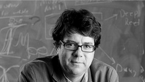
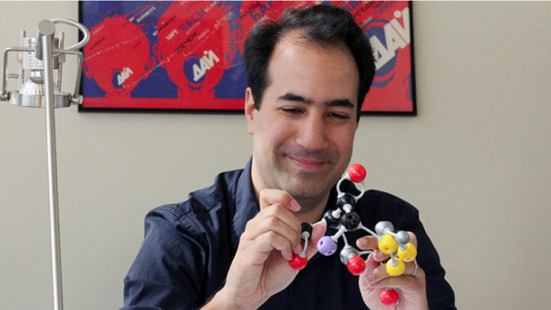
Assistant Professor, UCSD School of Medicine President/CEO, Institute for Brain and Society Affiliate, Krasnow Institute for Advanced Studies
Dr. Annese’s primary goal in the field of neuroscience is to conduct research that is open to public engagement and promotes the highest standards in data sharing and collaboration within the scientific community.
Institute website: Institute for Brain and Society Faculty website: UCSD School of Medicine
Email: jannese@ucsd.edu Office Phone: 858-822-4465 Lab Phone: 858-534-3177
Address: University of California, San Diego 3510 Dunhill Street San Diego, CA 92121
In 2005 Dr. Annese founded The Brain Observatory and in 2009 the laboratory was charged with the postmortem brain examination of one of the most famous medical cases in the history of neurology. The project evolved into the Digital Brain Library a novel collection of neurological and biographical data from medical patients and ordinary healthy individuals who have chosen to donate the brain to the project. The preservation and curation of their brain images and stories will help physicians and researchers understand the relationship between the brain, behavior and susceptibility to disease.
University of Rome ‘La Sapienza’ Rome, Italy B.A. and M.S. Biological Sciences
University College London London M.S., M.Phil. Neurosciences
Dartmouth College Hanover, NH Ph.D. Cognitive Neuroscience
OnAir Post: Jacopo Annese, PhD – UCSD

Associate Professor, The Vivian L. Smith Department of Neurosurgery Principal Investigator, Neuroimaging and Electrophysiology Lab
Dr. Tandon has co-authored many studies, which have been published in NeuroImage, Journal of Neurosurgery, Clinical Neurosurgery and Human Brain Mapping. His current research studies include “The localization of eloquent cortex using functional imaging and using diffusion tensor imaging tractography” and “The electrophysiological characteristics of language regions.”
Department webpage: med.uth.edu/neurosurgery/faculty/nitin-tandon/
Neuroimaging and Electrophysiology Lab website: tandonlab.org/
Email: Adriana.Garza@uth.tmc.edu (Administrative Assistant)
Phone: 713.704.7100
Address: UNIVERSITY OF TEXAS HEALTH SCIENCE CENTER AT HOUSTON 6431 FANNIN STREET, SUITE MSB G550D HOUSTON, TX 77030
OnAir Post: Nitin Tandon, MD – UT Health

USTAR Assistant Professor, Elect & Computer Engineering, University of Utah Associate Professor, Elect & Computer Engineering, University of Utah
Dr. Menon’s research lies at the intersection of optics and nanotechnology, with special foci on extending the spatial resolution of optics to the nanoscale, and applications of optics in energy. In addition to gaining a deep understanding of the basic physics of the behavior of light and matter at the nanoscale, our research is driven by many exciting applications.
Utah webpage: faculty.utah.edu/u0676529-Rajesh_Menon/research/index.hml
Nano Institute of Utah page: nanoinstitute.utah.edu/profiles/menon.php
University of Utah Neuroscience Initiative: http://brain2015.onair.cc/university-of-utah-neuroscience-initiative/
Email: rmenon@eng.utah.edu
Phone: 801-585-1058
Address: The University of Utah 50 S. Central Campus Dr. Rm 3280 Joseph F. Merrill Engineering Building Salt Lake City, UT 84112
OnAir Post: Rajesh Menon, PhD – Utah

Associate Professor of Bioengineering and BioMedical Engineering, Georgia Tech Principal Investigator, Precision Biosystems Laboratory Facilitator of the Invention Studio
Forest conducts research on miniaturized, high-throughput robotic instrumentation to advance neuroscience and genetic science, working at the intersection of bioMEMS, precision machine design, optics, and microfabrication. Prior to Georgia Tech, he was a research fellow in Genetics at Harvard Medical School.
Webpage: me.gatech.edu/faculty/forest Neuro@Tech Brain Initiative Grant
Email: craig.forest@me.gatech.edu Phone: 404-385-7645 Address: IBB Building, Room 1310
Dr. Craig Forest joined the Woodruff School of Mechanical Engineering as an Assistant Professor in August 2008. Since then he has established a research program focused on the creation and application of miniaturized, high-throughput robotic instrumentation to advance biomolecular science, along with the fundamental engineering that makes such instrumentation possible. Dr. Forest’s laboratory works at the intersection of bioMEMS, machine design, signal processing, optics, and manufacturing at the frontiers of the emerging bio-nano field. The development of instruments that can load, manipulate, and measure many biological samples at the resolution of single cells simultaneously with better accuracy and reliability than current approaches opens the door to essential, comprehensive biological system studies.
“New directions in science are ...
OnAir Post: Craig Forest, PhD – Ga. Tech
Professor of biology and biological engineering at Caltech Director, Tsao Lab
Doris Ying Tsao is a systems neuroscientist interested in the neural mechanisms underlying primate vision i.e. how visual objects are represented in the brain, and how these representations are used to guide behavior. She is investigating mechanisms at multiple stages in the visual hierarchy. Techniques we use include: electrophysiology, fMRI, electrical microstimulation, anatomical tracing, psychophysics, and mathematical modeling.
Website: cns.caltech.edu/people/faculty/tsao Lab: brain2015.onair.cc/tsao-lab/
Email: dortsao@caltech.edu Phone: 626-395-1702 Address: 34 Broad
Harvard University PhD 2002 Neuroscience (Advisor: Margaret Livingstone)
California Institute of Technology BS 1996 Biology and Math
I am a systems neuroscientist interested in the neural mechanisms underlying primate vision. The central problem I want to understand is how visual objects are represented in the brain, and how these representations are used to guide behavior. To address this, my lab is investigating mechanisms at multiple stages in the visual hierarchy, from early processes for segmenting visual input into discrete objects, to midand high-level perceptual processes for assigning meaningful identity to specific objects, to processes by which these perceptual representations govern behavior. Techniques we use include: electrophysiology, fMRI, electrical microstimulation, anatomical tracing, psychophysics, and mathematical modeling.
Ohayon S, Grimaldi P, Schweers N, Tsao D. Saccade modulation evoked by ...
OnAir Post: Doris Ying Tsao, PhD – Caltech
Professor of Brain & Cognitive Sciences and Biology, MIT Neuroscience Principal Investigator, Nedivi Lab
The Nedivi lab, part of the Picower Institute for Learning and Memory, studies the cellular mechanisms that underlie activity-dependent plasticity in the developing and adult brain through studies of neuronal structural dynamics, identification of the participating genes, and characterization of the proteins they encode.
Department of Brain and Cognitive Sciences page: biology.mit.edu/people/elly_nedivi
Picower Institute for Learning and Memory page: picower.mit.edu/Faculty/
Lab page: web.mit.edu/nedivi-lab/
MIT Neuroscience: neuroscience.onair.cc/mit-neuroscience/
Email: nedivi@mit.edu
Phone: 617-253-2344
Address: Room 46-3239
Elly Nedivi received her Ph.D. in Neuroscience from Stanford University Medical School and completed her postdoctoral training at The Weizmann Institute in Israel. In 1998, after two years at Cold Spring Harbor Laboratory, she joined the faculty of the Department of Brain and Cognitive Sciences and the Picower Institute for Learning and Memory at MIT. She also has an appointment in the Department of Biology at MIT.
Julie Martin Mid-Career Award in Aging Research
Edgerly Innovation Fund Award
Dean’s Education and Student Advising Award
Sloan Research Fellow
NSF POWRE Award
Ellison New Scholar Award
To understand the cellular mechanisms that underlie activity-dependent plasticity in the developing and adult brain, we are identifying and characterizing the participating genes and the function of ...
OnAir Post: Elly Nedivi, PhD – MIT
Associate Professor of Biology and Applied Physics, Stanford HHMI Investigator Principal Investigator, Schnizer Group
Dr. Schnitzer has longstanding interests in neural circuit dynamics and optical imaging focusing on: the development and application of fiber-optic, micro-optic, and nanophotonic imaging techniques for studies of learning and memory; in vivo fluorescence imaging and behavioral studies of hippocampal-dependent cognition and learning; and development of high-throughput, massively parallel imaging techniques for studying brain function in Drosophila.
Webpage: stanford.edu/dept/app-physics/cgi-bin/person/schnitzer-mark-j/ Stanford School of Medicine webpage: med.stanford.edu/profiles/mark-schnitzer Stanford Neurosciencs Institute Brain Initiative Grant
Email: mschnitz@stanford.edu Phone: 650) 723-4027 Address: James H. Clark Center – Room W080 318 Campus Drive Stanford, CA 94305
Harvard University Cambridge, MA A.B. summa cum laude 1988-1992 Physics
Cambridge University Cambridge, UK Certificate 1992-1993 Mathematics Princeton University Princeton, NJ M.A. 1993-1994 Physics
Princeton University Princeton, NJ Ph.D. 1994-1999 Physics (advisor: Prof. Steven M. Block)
2008-present Investigator, Howard Hughes Medical Institute; Stanford University.
2006-present Janelia Farm Research Campus, Howard Hughes Medical Institute, Scientific Visitor Program, Ashburn VA.
2003-present Assistant Professor, Dept. of Applied Physics and Dept. of Biological Sciences; Faculty Member, Neuroscience Program, Biophysics Program, Stanford Univ., Stanford, CA.
1999-2003 Member of Technical Staff, Physical Sciences Laboratory, Biological Computation Research Department, Bell Laboratories, Lucent Technologies, Murray Hill, NJ.
1994-1999 Ph.D. Research, with Steven M. Block, Dept. of Molecular Biology, ...
OnAir Post: Mark J Schnitzer, PhD – Stanford
Professor, Biological Sciences and Neuroscience and Co-Director, Kavli Institute for Brain Science at Columbia University Member, Multi-Council Working Group (BRAIN Initiative) Member, Advisory Committee to the Director (NIH)
Dr. Yuste has pioneered the application of imaging techniques, such as calcium imaging of neuronal circuits, two-photon imaging, photostimulation using caged compounds and holographic spatial light modulation microscopy.
Columbia/Kavli Webpage: kavli.columbia.edu/leadership/yuste Lab Webpage: columbia.edu/cu/biology/faculty/yuste/ Allen Institute Webpage: alleninstitute.org/our-institute/advisors/profiles/rafael-yuste/ Twitter: @yusterafa
Email: rafaelyuste@columbia.edu Phone: 212-854-5023 Address: 901 NWC Building 550 West 120th Street New York, NY 10027
The goal of Dr. Yuste’s research is to understand the function of the cortical microcircuit. The cortex constitutes the larger part of the brain in mammals. In humans it is the primary site of mental functions like perception, memory, control of voluntary movements, imagination, language and music. No accepted unitary theory of cortical function exists yet; nevertheless, the basic cortical microcircuitry develops in stereotyped fashion, is similar in different cortical areas and in different species, and has apparently not changed much in evolution since its appearance. At the same time, the cortex participates in apparently widely different computational tasks, resembling a “Turing machine”. Because of this, it is conceivable that a “canonical” cortical microcircuit may exist and implement a relatively simple, and flexible, computation.
We pursue the reverse-engineering of the ...
OnAir Post: Rafael Yuste, MD, PhD – Columbia
Principal Investigator: Mark J Schnitzer Stanford Neurosciencs Institute
The Schnitzer Group has three major research efforts: Development and application of fiber-optic, micro-optic, and nanophotonic imaging techniques for studies of learning and memory in behaving mice and for clinical uses in humans; In vivo fluorescence imaging and behavioral studies of hippocampal-dependent cognition and learning; and Development of high-throughput, massively parallel imaging techniques for studying brain function in large numbers of Drosophila concurrently.
Website: http://pyramidal.stanford.edu/index.html Brain Initiative Grant
Email: schnizerlab@gmail.com Address: James H. Clark Center – Room W080 318 Campus Drive Stanford, CA 94305
Dr. Schnitzer has longstanding interests in neural circuit dynamics and optical imaging, and his laboratory has three major research efforts:
In vivo fluorescence imaging and behavioral studies of cerebellar-dependent motor control and motor learning. Development and application of fiber-optic fluorescence microendoscopy imaging techniques for studies of learning and memory in behaving mice and for clinical uses in humans. Development of high-throughput, massively parallel imaging techniques for studying brain function in large numbers of Drosophila concurrently.The long-term goal of our research is to advance experimental paradigms for understanding normal cognitive and disease processes at the level of neural circuits, with emphasis on learning and memory processes. By contrast, much current research on learning and memory concentrates on ...
OnAir Post: Schnitzer Group – Stanford
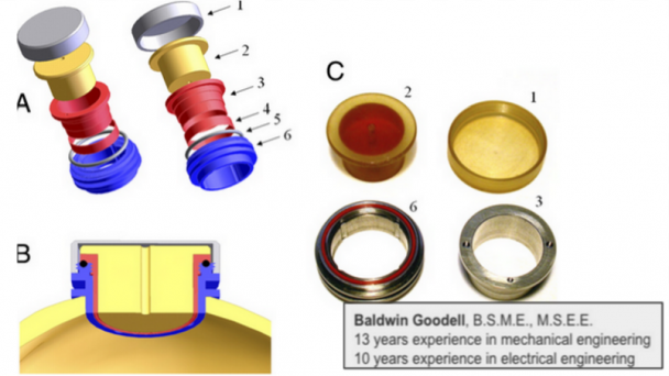
Principal Investigator: Gray Matter Research
Goodell and his company Gray Matter Research focuses on Microdrive Systems and Recording Chamber Systems. For the BRAIN Initiative, Goodell and his colleagues aim to develop optrodes, which are implantable columns of lights and wires for simultaneous electrical recording of neurons and delivery of light flashes to multiple brain areas.
Email: baldwin@graymatter-research.com Phone: (406) 672-1915 Address: Gray Matter Research 920 Technology Blvd. Suite 106 Bozeman, MT 59718
OnAir Post: Albert Baldwin Goodell – Gray Matter Research
Professor of Biomedical Engineering, Washington University Director, Optical Imaging Laboratory
His lab reported the first functional photoacoustic tomography, 3D photoacoustic microscopy (PAM), optical-resolution PAM, photoacoustic Doppler effect, photoacoustic reporter gene imaging, microwave-induced thermoacoustic tomography, the universal photoacoustic reconstruction algorithm, frequency-swept ultrasound-modulated optical tomography, time-reversed ultrasonically encoded (TRUE) optical focusing, sonoluminescence tomography, Mueller-matrix optical coherence tomography, and optical coherence computed tomography.
Webpage: bme.wustl.edu/people/Pages/faculty-bio.aspx?faculty=19 Washington University Neuroscience Program BRAIN Initiative Grant – “Fast High-Resolution Deep Photoacoustic Tomography of Action Potentials in Brains”
Email: lhwang@biomed.wustl.edu Phone: (314) 935-6152 Address: One Brookings Drive Campus Box 1097 Whitaker Hall, Room 190D St. Louis, MO 63130
Lihong Wang earned his Ph.D. degree at Rice University, Houston, Texas under the tutelage of Robert Curl, Richard Smalley, and Frank Tittel and currently holds the Gene K. Beare Distinguished Professorship of Biomedical Engineering at Washington University in St. Louis.
His book entitled “Biomedical Optics: Principles and Imaging,” one of the first textbooks in the field, won the Joseph W. Goodman Book Writing Award. He also coauthored a book on polarization and edited the first book on photoacoustic tomography. Professor Wang has published more than 420 peer-reviewed articles in journals including Nature (Cover story), Science, PNAS, and PRL with an h-index of 93 (Google Scholar) and delivered over ...
OnAir Post: Lihong Wang, PhD – Washington U

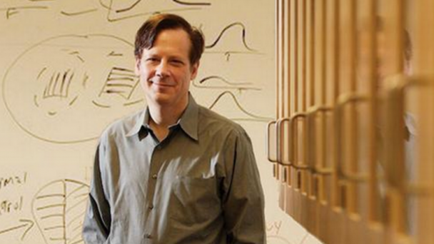
Assistant Professor of Biology in Boston University Department of Biology Assistant Professor, Biomedical Engineering Principal Investigator, Gardner Lab
Gardner studies the mechanisms of temporal sequence perception and production, focusing on vocal learning in songbirds.The song circuit produces stereotyped structure over a range of time-scales from milliseconds to tens of seconds. He also develops minimally invasive electrodes that provide stable neural recordings in behaving animals.
Webpage: bu.edu/bme/people/joint/gardner/ Lab: http://people.bu.edu/timothyg/index.html Brain Initiative Grant
Email: timothyg(at) bu.edu Phone: (347) 683-7642 Address: 24 Cummington Mall Room 402 Boston, MA 02215
PhD, Rockefeller University
Research interests include: Neural circuits, vocal learning, time-frequency analysis, brain-machine interfaces
The Gardner lab studies the mechanisms of temporal sequence perception and production, focussing on vocal learning in songbirds.
The song circuit produces stereotyped structure over a range of time-scales from milliseconds to tens of seconds. We ask how complex songs are assembled from elementary neural units. What are the relationships between patterns of neural activity on different time-scales?
The lab also studies information processing in auditory cortex, examining how auditory signals are transformed as they move from low to high level sensory areas. How are memories for temporal patterns formed?
To address these questions, we develop minimally invasive electrodes that provide stable neural recordings in behaving animals. We also develop high-resolution signal processing algorithms ...
OnAir Post: Tim Gardner, PhD – BU
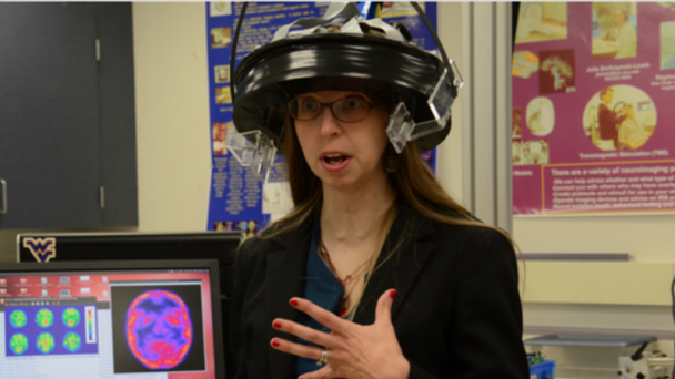
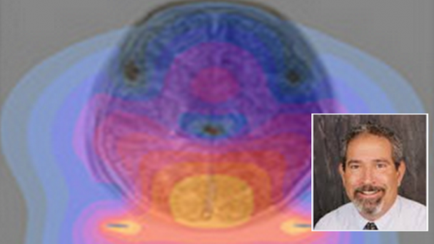
Research Assistant Professor, Department of Physiology Director, Brefczynski-Lewis Lab
Brefczynski-Lewis studies how we perceive people we love and people we don’t like, both famous and political, and how training in compassion can affect those perceptions. She is examining the neural and physiological correlates of the liked and disliked persons and how these change after training in compassion. Grudge forgiveness study: fMRI response to the face of the grudge person, as well as cardio and reactive measures will be tested before and after the intervention.
Webpage: directory.hsc.wvu.edu/UserDetails/36369 WVU Center for Neuroscience BRAIN Initiative Grant – “Imaging the Brain in Motion: The Ambulatory Micro-Dose, Wearable PET Brain Imager”
Email: jblewis@hsc.wvu.edu Phone: 304-293-6898 Address: Radiology Research One Medical Center Drive HSC South, PO Box 9236 Morgantown, WV 26506-9236
Medical College of Wisconsin, Doctor of Philosophy (PhD), Cell Biology, Neurobiology and Anatomy 1996 – 2004
Lawrence University, Bachelor of Arts (B.A.), Biology with Interdisciplinary Neuroscience 1993 – 1997
My research experience has been in studying the neural correlates of cognitive, affective and social processes. Specifically I have focused in recent years in examining the effects of training in compassion meditation and empathy on brain activation and behavior. I have a publication and a private grant in ...
OnAir Post: Julie Brefczynski-Lewis, PhD – WVU
Professor, University of Minnesota Center for Magnetic Resonance Research
Garwood focus has been on developing cutting-edge MRI and MR spectroscopy techniques and on exploiting them in studies of tissue function, metabolism, and microstructure. An emphasis has been on identifying and validating quantitative metrics to assess normal and disease states non-invasively with imaging, and on applying them to learn about metabolism, hemodynamics, and tissue micro-environment.
Webpage: cmrr.umn.edu/facultystaff/gar.shtml Institute for Translational Neuroscience BRAIN Initiative Grant
Email: gar@cmrr.umn.edu Phone: 612-626-2001 Address: 1-211B CMRR
For the past 26 years, researchers in the Garwood laboratory have had a focus on developing cutting-edge MRI and MR spectroscopy techniques and on exploiting them in studies of tissue function, metabolism, and microstructure. An emphasis has been on identifying and validating quantitative metrics to assess normal and disease states non-invasively with imaging, and on applying them to learn about metabolism, hemodynamics, and tissue micro-environment. On the technical side, the Garwood group has recently made a significant advancement in the way MRI is performed – a technique called SWIFT. SWIFT exploits time-shared RF excitation and acquisition to preserve signals from water molecules possessing extremely short transverse relaxation times, T2 and T2*. With SWIFT, ...
OnAir Post: Michael Garwood, PhD – Minnesota

Professor, Psychiatry and Biobehavioral Sciences, UC Los Angeles Director, X. William Yang Research Group
Yang is interested in using the mouse molecular genetic approach to study the pathogenesis of neurodegenerative diseases. One recurring theme in neurodegenerative diseases is that a widely expressed mutant protein can cause highly selective degeneration of a subset of neurons. The pathogenesis of such selective neurodegeneration remains unclear. Currently, we are focusing on Huntington’s disease (HD) to study the molecular and cellular mechanisms underlying the disease.
Webpage: bioscience.ucla.edu/faculty/x-william-yang UCLA Neuroscience BRAIN Initiative Grant– “Novel Genetic Strategy for Sparse Labeling and Manipulation of Mammalian Neurons”
Email: xwyang@mednet.ucla.edu Phone: 310-267-2761 Address: 695 Charles Young Drive, #3309 Los Angeles, CA 90095 695 Charles Young Drive, Gonda 3506B Los Angeles, CA 90095
Dr. X. William Yang is a professor in the Department of Psychiatry & Biobehavioral Sciences at David Geffen School of Medicine at UCLA. He is also a member of the Center for Neurobehavioral Genetics at Semel Institute for Neuroscience & Human Behaviors, and a member of the Brain Research Institute at UCLA. He has served as a regular member at the NIH’s Cell Death in Neurodegeneration (CDIN) Study Section, a Scientific Advisory Board member of the Hereditary Disease Foundation, and a faculty member for Faculty 1000 Medicine?s Neurogenetics ...
OnAir Post: X. William Yang, MD/PhD – UCLA

Professor of Neurosurgery and Physiology, Mayo Clinic
The research interests of Kendall H. Lee, M.D., Ph.D., are to develop deep brain stimulation (DBS) for the treatment of Parkinson’s disease, tremor, depression, obsessive-compulsive disorder and epilepsy. Dr. Lee is fascinated with the possibility of combining sophisticated electrophysiological recordings with miniaturized analytical elements (microprocessors) to augment or repair disrupted function of the brain.
Webpage: http://www.mayo.edu/research/faculty/lee-kendall-h-m-d-ph-d/bio-00027489 Department of Physiology and Biomedical Engineering: mayo.edu/research/department-physiology-biomedical-engineering Brain Initiative Grant
Email: Lee.Kendall@mayo.edu Address: Joseph Building 4-184W 200 First St. SW Rochester, MN 55905
Chief Resident – Neurosurgery Dartmouth Hitchcock Medical Center
Resident – Neurosurgery Dartmouth Hitchcock Medical Center
Internship – General Surgery Dartmouth Hitchcock Medical Center
Resident – Neurology Partners’ Neurology Program, Harvard Medical School
Internship – Internal Medicine Hospital of St. Raphael, Yale University School of Medicine
PhD Department of Neurobiology, Yale University Graduate School
MD Yale University Graduate School
M. PhilYale University Graduate School
BA – Major-Biology/Minor-Philosophy University of Colorado, Denver
The research interests of Kendall H. Lee, M.D., Ph.D., are to develop deep brain stimulation (DBS) for the treatment of Parkinson’s disease, tremor, depression, obsessive-compulsive disorder and epilepsy. Dr. Lee is fascinated with the possibility of combining sophisticated electrophysiological recordings with miniaturized analytical elements (microprocessors) to augment or repair disrupted function of the brain.
OnAir Post: Kendall H Lee, MD/PhD – Mayo
Professor & Chair: Anatomy & Neurobiology, Physiology & Biophysics, and Neurobiology & Behavior Director, Soltesz Lab
Research Focus: Working to understand: traumatic brain injury, post-traumatic epilepsy, fever-induced (Febrile) seizures in childhood, learning and memory deficits.
Scientific Focus: functions, development and plasticity of hippocampal interneuronal networks. Physiological basis of hyperexcitability. Mechanisms of selective neuronal vulnerability.
Webpage: anatomy.uci.edu/soltesz.html UC Irvine Neuroscience Brain Initiative Grant – “Towards a Complete Description of the Circuitry Underlying Memory replay”
Email: isoltesz@uci.edu Phone: 949-824-3957 and 3967 Address: Dep’t Anatomy & Neurobiology 117 Irvine Hall School of Medicine University of California Irvine, California 92697-1280
1983-1988
Diploma in Biology L. Eotvos University, Budapest, Hungary1988-1989
Ph.D., Comparative Physiology L. Eotvos University, Budapest, Hungary1989-1990
Postdoctoral Research Fellow, MRC Anatomical Neuropharmacology Unit Oxford University, England1990-1991
Postdoctoral Researcher, Dept of Visual Science, Institute of Opthalmology University of London, U.K.1991-1992
Post-graduate Postdoctoral Researcher, Centre de Recherche en Neurobiologie Université Laval, Quebec1992-1993
Postdoctoral Researcher, Dept of Neurology and Neurological Sciences Stanford University, Palo Alto, California1993-1995
Postdoctoral Researcher, Dept of Anesthesiology and Pain Management UT Southwestern, Dallas, Texas1995-1999
Assistant Professor, Dept.’s of Anatomy & Neurobiology and Physiology & Biophysics University of California, Irvine, California1999-2003
Associate Professor, Dept.’s of Anatomy & Neurobiology and Physiology & Biophysics University of California, Irvine, California2002-2003
Associate Professor, Dept. of Neurobiology & Behavior University of California, Irvine, California2001-present
Fellow, Center for the Neurobiology of Learning & Memory University of California, Irvine, California2003-present
Professor, ...OnAir Post: Ivan Soltesz, PhD – UC Irvine

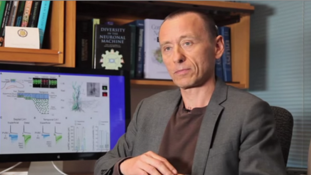
Core Faculty, Program in Biological Sciences, UCSF Physiology Department Director: Frank Laboratory
Frank’s research interests center around learning and spatial coding in the hippocampal-cortical circuit. Frank is interested in understanding the neural correlates of learning and memory. In particular, his laboratory focuses on the circuitry of the hippocampus and adjacent regions. His goal is to examine the relationships among neural firing patterns, behavior, and anatomy to understand how the brain uses and stores information.
Webpage: keck.ucsf.edu/physio/people/frankl.html#research UCSF Neuroscience Brain Initiative Grant
Email: loren@phy.ucsf.edu Phone: 415-502-6317 Address: UCSF 513 Parnassus Box 0444 San Francisco, CA 94143-0444
The ability to use experience to guide behavior (to learn) is one of the central functions of the brain. We are interested in understanding the neural correlates of learning and memory. In particular, our laboratory focuses on the circuitry of the hippocampus and adjacent regions. Our goal is to examine the relationships among neural firing patterns, behavior, and anatomy to understand how the brain uses and stores information. Ultimately we should be able to generate accurate computational models of learning to both test hypotheses concerning hippocampal-cortical interactions and to generate new predictions that can be tested experimentally.
The hippocampal formation has a unique anatomical organization in that the connectivity between adjacent hippocampal regions is ...
OnAir Post: Loren M Frank, PhD – UCSF
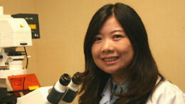
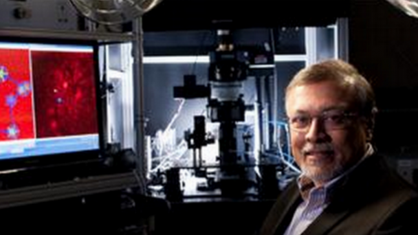
Assistant Professor, Department of Biochemistry and Molecular Medicine, UC Davis Director, Tian Lab
The goal of Tian’s research is to invent new molecular tools for analyzing and engineering functional neural circuits. We also leverage these tools, combined with optical imaging techniques, to study molecular mechanisms of neurological disorders at system level and to empower searching for novel therapeutic treatments.
Webpage: ucdmc.ucdavis.edu/biochem/faculty/tian/ UC Davis Neuroscience Brain Initiative Grant
Email: lintian@ucdavis.edu Phone: (916) 734-8070 Address: 2352 Oak Park Research Building Sacramento Campus
I was born and raised in China. After graduating from University of Science and Technology of China, I joined a interdisciplinary PhD program at Northwestern University, where I studied the mechanisms of protein processing via ubiquitin-proteasome pathway in Dr. Andreas Matouschek’s lab. I then moved to HHMI Janelia Farm as a postdoc. The highly collaborative environment at Janelia resulted in my multidisciplinary training under three principle investigators, Dr. Loren Looger, Dr. Karel Svoboda and Dr. Luke Lavis. There, my research focused on engineering optical probes for monitoring and controlling neural circuitry in living behaving animal. These new imaging techniques have greatly impacted the field of neuroscience, facilitating new types of biological experiments performed to address previously intractable questions. One indication of the impact of this ...
OnAir Post: Lin Tian, PhD – UC Davis

Professor of Child Neurology and Mental Retardation, Harvard Medical School Unit Chief, Pediatric Neurology, Massachusetts General Hospital Director, Pediatric Epilepsy Research Lab
Staley focuses on neuronal ion transport and the spread of activity in neural networks. Research interests include epilepsy, synaptic physiology, and neural network activity. Research techniques used: single cell electrophysiology, in vivo radiotelemetry, ion-sensitive fluorescent imaging of ion transport and neural network activity, computer modeling.
Webpage: massgeneral.org/neurology/researcher_profiles/staley_kevin Neuroscience@Harvard BRAIN Initiative grant
Email: staley.kevin@mgh.harvard.edu Clinic Phone: 617-724-6400 Address: Kevin J. Staley, MD Massachusetts General Hospital 114 16th Street Charlestown, MA 02129
Joseph P. and Rose F. Kennedy Professor of Neurology, Harvard Medical School
Unit Chief, Pediatric Neurology, Mass General Hospital Department of Neurology
Kevin Staley received his MD degree from the University of California, San Diego. He completed his postdoctoral research training at Stanford University School of Medicine. Dr Staley studies neuronal ion transport in neonatal seizures and neural network dysfunction in epilepsy. He has served as Chair of the Investigator’s Workshop Committee and the Research and Training Committee of the American Epilepsy Society, as Chair of the Research Council of the Epilepsy Foundation of America, as co-chair of the inaugural Gordon Conference on Mechanisms of Epilepsy and Neuronal Synchronization, and as an Associate Editor for the Journal of ...
OnAir Post: Kevin J. Staley, MD – Harvard Med
Professor of Cellular and Molecular Physiology and of Neurobiology, Yale University Fellow, John B. Pierce Laboratory
Dr Pieribone is developing genetically encoded fluorescent probes of membrane electrical potential. These probes allow one to use optical instruments (microscopes) to monitor the electrical activity of neurons. He has also engineered miniature imaging systems that can be head mounted on mammels and allow mobile recording of neuronal activity.
Webpage: medicine.yale.edu/bbs/people/vincent_pieribone-3 Fluorogenetic Voltage Sensors website: fluorogenetic-voltage-sensors.org/ Yale Neuroscience Brain Initiative Grant
Email: vincent.pieribone@yale.edu Phone: (203) 562-9901 x214 Address: The John B. Pierce Laboratory 290 Congress Avenue New Haven, CT 06519
B.A. New York University, Washington Square University College, Biology and Chemistry, 1986
Ph.D. New York University, Graduate School, Washington Square, Neurobiology, 1992
Research Associate, The American Museum of Natural History Scientific Board of Directors, Mystic Aquarium and Center for Exploration
The brain uses complex and highly parallel computational paradigms to process sensory information, create and retrieve memories, and execute motor actions. The unit of this computing network is the neuron and its attendant synaptic connections. The structure and physiology of the brain makes direct study of these structures in the living organisms very difficult – neurons and synapses are tiny, very delicate, and tightly packed. Our laboratory is dedicated to the study of how neuronal ...
OnAir Post: Vincent Allen Pieribone, PhD – Yale
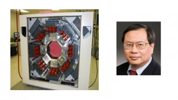
Professor, Johns Hopkins Medicine Department of Radiology and Radiological Science Radiology Vice Chair, Research Administration and Training Director Section of High Resolution Brain PET Imaging, Division of Nuclear Medicine
Dr. Wong has used PET scanning to uncover key insights into brain chemistry and to identify receptors for the major neurotransmitters. He oversaw the first dopamine PET receptor imaging in human beings; led the first study suggesting D2 dopamine receptors in schizophrenia, and how dopamine is transported in and out of cells.
Webpage: neuroscience.jhu.edu/resources/directory/faculty/dean-f.-wong-m.d.-ph.d/ Brain Initiative Grant
Email: dfwong@jhmi.edu Phone: 410-955-8433 Address: Johns Hopkins University School of Medicine Department of Neuroscience 1003 Wood Basic Science Building 725 N. Wolfe St. Baltimore, MD 21205
MD (University of Toronto)
PhD (Johns Hopkins University)
The use of novel methods in positron emission tomography (PET) and single photon emission computed tomography (SPECT) have, in the past few decades, been used to study a wide variety of neuropsychiatric illness, basic brain chemistry and pharmacology. Our focus is on the design, development and application of radiopharmaceuticals imaged PET and SPECT for the study of in vivo brain chemistry. Our research extends from collaborations in basic chemistry ...
OnAir Post: Dean Foster Wong, MD/PhD – JHU
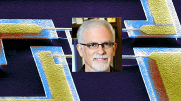
Professor of Physics, Applied Physics, and Bioengineering, CalTech Division of Engineering and Applied Sciences Director, Roukes Group
Roukes research activities are currently focused on developing advanced nanodevices, engineering them into complex systems, and using them to enable fundamental problems in neuroscience and proteomics. A continuing thread in theoretical and experimental investigations focuses on fundamental properties of nanomechanical systems.
Lab webpage: caltech.edu/people/3185/profile Division webpage: nano.caltech.edu/people/roukes Caltech Neuroscience Brain Initiative Grant
Email: roukescaltech.edu Phone: 626-395-2916 Address: MC 149-33Pasadena, CA 91125
B.A., University of California (Santa Cruz), 1978; Ph.D., Cornell University, 1985. Associate Professor, Caltech, 1992-96; Professor of Physics, 1996-2002; Professor of Physics, Applied Physics, and Bioengineering, 2002-11; Abbey Professor, 2011-; Director, Kavli Nanoscience Institute, 2004-06; Co-Director, 2008-2013.
Professor Roukes’s research focuses on nanobiotechnology, nanotechnology, nanoscale physics, nanoscale and molecular mechanics.
nanobiotechnology, nanotechnology, nanoscale physics, nanoscale and molecular mechanics
The Kavli Nanoscience Institute, Center for the Physics of Information
OnAir Post: Michael Roukes, PhD – CalTech

Professor of Radiology, Neurobiology, Psychiatry and Behavioral Science and Biomedical Engineering Director, Duke-UNC Brain Imaging and Analysis Center
Allen Song’s research interests focus on the acquisition methodology, processing strategies and contrast mechanism for functional MRI. Additional interests include the application of innovative fMRI acqusition and analysis methods to study functional neuroanatomy.
Webpage: biac.duke.edu/people/asong Duke Institute for Brain Sciences Brain Initiative Grant
Email: allen.song@duke.edu Phone: (919) 684-1215 Address: Duke-UNC Brain Imaging and Analysis Center Duke University Hock Plaza, Suite 501 2424 Erwin Road Durham, NC 27705
Ph. D., 1995, Medical College of Wisconsin (Biophysics)
Post-Doctoral Fellowship, Lab of Brain and Cognition, NIH
The acquisition methodology, processing strategies and contrast mechanism for functional MRI. Additional interests include the application of innovative fMRI acqusition and analysis methods to study functional neuroanatomy.
The research in this lab is concerned with the advancement of fMRI data acquisition methods that includes the development of real-time imaging using echo-planar and spiral data acquisition with high-order shimming control, development of robust and reliable single-shot image acquisition methods and optimization of the acquisition methods for improved functional sensitivity and specificity.
Our lab is also focused on understanding the contrast mechanism of the functional MRI, which includes the source localization of the functional signal using the blood ...
OnAir Post: Allen Song, PhD – Duke
Zarem Professor of Bioengineering, Caltech Neuroscience Director, Dickinson Lab
The aim of Dickinson’s research is to elucidate the means by which flies accomplish their aerodynamic feats. A rigorous mechanistic description of flight requires an integration of biology, engineering, fluid mechanics, and control theory. The long term goal, however, is not simply to understand the material basis of insect flight, but to develop its study into a model that can provide insight to the behavior and robustness of complex systems in general.
Webpage: eas.caltech.edu/people Lab: http://depts.washington.edu/flyarama/ TEDx video: ted.com/talks/michael_dickinson_how_a_fly_flies Wikipedia Entry: wiki/Michael_Dickinson BRAIN Initiative grant
Email: flymancaltech.edu Phone: 626-395-5775 Address: The California Institute of Technology Mail Code 216-76 Pasadena, CA 91125
from Wikipedia page
Michael H. Dickinson (born 1963) is an American fly bioengineer and neuroscientist, and Zarem Professor of Biology and Bioengineering at the California Institute of Technology. He studies Drosophila flight control systems and sensory processing.
He graduated from Brown University with a B.S. in 1984, and from University of Washington with a Ph.D. in 1989. He was previously part of the faculty at the University of Chicago, the University of California, Berkeley, and the University of Washington.
He is a Monitoring Editor at the Journal of Experimental Biology.He was a course director of the Neural Systems and ...
OnAir Post: Michael Dickinson, PhD – Caltech

Professor at Cold Springs Harbor Laboratory & HHMI Investigator Principal Investigator, Hannon Lab
Greg Hannon explores the processes that cells use to turn genes on and off. My work is focused on understanding a relatively new class of cellular pathways, governed by molecules known as small RNAs, that control gene activation and repression. Our studies of small-RNA biology in early development provide insights into human evolution, diversity, and diseases such as cancer.
CSHL Website: cshl.edu/Faculty/Gregory-Hannon.html HHMI webpage: hhmi.org/scientists/gregory-j-hannon Lab: hannonlab.cshl.edu/index.html
Email: hannon@cshl.edu Phone: (516) 367-8455 Address: One Bungtown Road Cold Spring Harbor, NY 11724
Ph.D., Case Western Reserve University,1992
I explore the processes that cells use to turn genes on and off. My work is focused on understanding a relatively new class of cellular pathways, governed by molecules known as small RNAs, that control gene activation and repression. Our studies of small-RNA biology in early development provide insights into human evolution, diversity, and diseases such as cancer.
Gregory Hannon is a pioneer in the study of RNA interference (RNAi), a process in which double-stranded RNA molecules induce gene silencing. Hannon and colleagues have elucidated key elements of the RNAi machinery. During the past several years, the Hannon lab has focused on the ...
OnAir Post: Gregory Hannon, PhD – CSHL
Associate Professor of Biological Engineering with appointments in Brain and Cognitive Sciences and Nuclear Science and Engineering, MIT Neuroscience Associate member of the McGovern Institute Principal Investigator, Jasanoff Lab
Functional magnetic resonance imaging (fMRI) has revolutionized our understanding of the human brain, but the method is now approaching the limit of its capabilities. Alan Jasanoff hopes to break through this limit and to develop new technologies for imaging the molecular and cellular phenomena that underlie brain function.
McGovern Webpage: mcgovern.mit.edu/principal-investigators/alan-jasanoff
Lab page: mit.edu/~jasanofflab/
Department of Brain and Cognitive Sciences page: bcs.mit.edu/people/jasanoff.html
MIT Neuroscience: neuroscience.onair.cc/mit-neuroscience/
Email: jasanoff@mit.edu
Phone:617-452-2538
Address: MIT Rm. 16-561 | 77 Massachusetts Avenue | Cambridge, MA 02139
Alan Jasanoff is an associate member of the McGovern Institute and Associate Professor of Biological Engineering, with appointments in Brain and Cognitive Sciences and Nuclear Science and Engineering. He was awarded tenure in 2011. Prior to joining the MIT faculty, he was a Whitehead Fellow at the Whitehead Institute for Biomedical Research at MIT. He was named a Raymond and Beverly Sackler Foundation Scholar in 2004 and received the McKnight Technological Innovations in Neuroscience Award in 2006. Jasanoff was also a 2007 recipient of the Director’s New Innovator Award from the National Institutes of Health.
Pushing the frontiers of ...
OnAir Post: Alan Jasanoff, PhD – MIT
Doris and Don Berkey Professor of Neuroscience, Department of Brain and Cognitive Sciences, Massachusetts Institute of Technology Director, McGovern Institute for Brain Research and Desimone Lab
Robert Desimone studies the brain mechanisms that allow us to focus our attention on a specific task while filtering out irrelevant distractions. Our brains are constantly bombarded with sensory information. The ability to distinguish relevant information from irrelevant distractions is a critical skill, one that is impaired in many brain disorders.
McGovern Webpage: mcgovern.mit.edu/principal-investigators
Lab page: desimonelab.org/robert-desimone/
Department of Brain and Cognitive Sciences page: bcs.mit.edu/people/desimone
MIT Neuroscience: brain2015.onair.cc/mit-neuroscience/
E-mail: desimone@mit.edu
Phone: 617-324-2077
Address: MIT Bldg 46-3160 | 43 Vassar Street | Cambridge, MA 02139
Robert Desimone is Director of the McGovern Institute and Professor in the Brain and Cognitive Sciences Department. Prior to coming to MIT, he was Director of the NIMH Intramural Research Program, the largest mental health research center in the world. Desimone received his B.A. from Macalester College and his Ph.D. from Princeton University . He is a member of the National Academy of Sciences and the American Academy of Arts of Sciences, and a recipient of numerous awards, including the Troland Prize of the National Academy of Sciences, and the Golden Brain Award of ...
OnAir Post: Robert Desimone, PhD – MIT

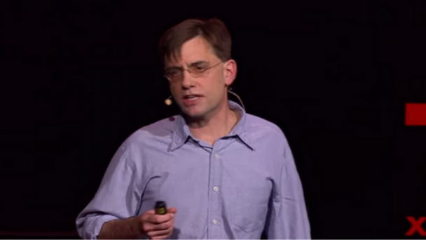
Associate Professor and AT&T Chair, MIT Media Lab and McGovern Institute, Departments of Biological Engineering and Brain and Cognitive Sciences Co-Director, MIT Center for Neurobiological Engineering Principal Investigator, Synthetic Biology Group
Ed Boyden develops new strategies for analyzing and engineering brain circuits to develop broadly applicable methodologies that reveal fundamental mechanisms of complex brain processes. A major goal of his current work is the development of technologies for controlling nerve cells using light.
Personal Website: edboyden.org/
McGovern Institute for Brain Research page: mcgovern.mit.edu/principal-investigators/ed-boyden
Lab Page: syntheticneurobiology.org/
Twitter: twitter.com/eboyden3
Wikipedia page: en.wikipedia.org/wiki/Edward_Boyden
E-mail: esb@media.mit.edu
Phone: (617) 324-3085
Address: Room E15-421 |20 Ames St. | Cambridge, MA 02139
From Lab Page
Ed Boyden is Associate Professor of Biological Engineering and Brain and Cognitive Sciences, at the MIT Media Lab and the MIT McGovern Institute. He leads the Synthetic Neurobiology Group, which develops tools for analyzing and engineering the circuits of the brain. These technologies, created often in interdisciplinary collaborations, include ‘optogenetic’ tools, which enable the activation and silencing of neural circuit elements with light, 3-D microfabricated neural interfaces that enable control and readout of neural activity, and robotic methods for automatically recording intracellular neural activity and performing single-cell analyses in the living brain. He has launched an ...
OnAir Post: Ed Boyden, PhD – MIT
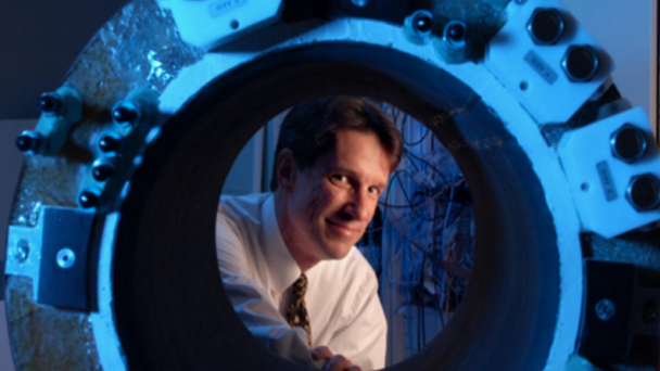
Adjunct professor of neuroscience at UC Berkely and of Radiology at UCSF Board Certified Diagnostic Radiologist and Neuroradiologist President, Advanced MRI Technologies (AMRIT)
Dr. Feinberg is an internationally recognized expert on Magnetic Resonance Imaging (MRI), with numerous publications and research studies to his credit. In addition, he holds many patents in MRI technology.
Redwood Regional Medical Group webpage: rrmginc.com/physicians Helen Wills Neuroscience Institute Brain Initiative Grant
Email: david.feinberg@advancedmri.com Phone: 707-829-2933 Address: Advanced MRI Technologies 652 Petaluma Ave, Suite J Sebastopol, CA 95472
Dr. Feinberg completed his B.A., M.S. and Ph.D., at the University of California, Berkeley. After completing his Ph.D, Dr. Feinberg attended the University of Miami, School of Medicine’s ‘Ph.D to M.D’ medical program. He remained on the east coast for his internship and residency at Brigham and Womens Hospital (Harvard Medical School), and at NYU Medical Center. He has a fellowship in Neuroradiology from Washington University. He received his board certification in Diagnostic Radiology in 1997.
Dr. Feinberg is an internationally recognized expert on Magnetic Resonance Imaging (MRI), with numerous publications and research studies to his credit. In addition, he holds many patents in MRI technology.
In his spare time he enjoys cycling, hiking and traveling.
From UC Berkely News 9/30/15
David Feinberg, a UC ...
OnAir Post: David Feinberg, MD/PhD – UCSF
Professor of Neuroscience, MIT Department of Brain and Cognitive Sciences Director, Simons Center for the Social Brain Principal Investigator, Laboratory of Mriganka Sur
Dr. Sur studies the organization, development and plasticity of the cerebral cortex of the brain using experimental and theoretical approaches. He has discovered fundamental principles by which networks of the cerebral cortex are wired during development and change dynamically during learning.
Webpage: web.mit.edu/msur/www/profile.html Simons Center for the Social Brain website: web.mit.edu/scsb/ MIT Neuroscience Brain Initiative Grant
Email: msur@mit.edu Phone: 617.253.8785 Address: 43 Vassar St. 46-6227 Cambridge, MA, 02139
Dr. Mriganka Sur is the Paul E. and Lilah Newton Professor of Neuroscience and Director of the Simons Center for the Social Brain at MIT, which he founded after 15 years as head of the MIT Department of Brain and Cognitive Sciences. Dr. Sur studies the organization, development and plasticity of the cerebral cortex of the brain using experimental and theoretical approaches. He has discovered fundamental principles by which networks of the cerebral cortex are wired during development and change dynamically during learning. His laboratory has identified gene networks underlying cortical plasticity, and pioneered high resolution imaging methods to study cells, synapses and circuits of the intact brain. Recently, his group has demonstrated novel mechanisms underlying disorders of brain ...
OnAir Post: Mriganka Sur, PhD – MIT

Research Scientist Head of MIT Genetic Neuroengineering Group
Research interests: viral vector engineering, synthetic biology. Engineering genetic tools for neuroscience.
LinkedIn Webpage: linkedin.com/in/ianwickersham MIT Neuroscience Brain Initiative Grant
Email: wickersham@mit.edu
Ian obtained a PhD from UCSD, where he developed new retrograde viral technologies for cell-targetable transsynaptic circuit tracing. After a postdoctoral fellowship in MIT Brain and Cognitive Science, he joined the Synthetic Neurobiology group as a research scientist to develop new integrative cell and circuit analysis methods. He then went on to launch the MIT Genetic Neuroengineering Group.
Ian is eveloping new integrative cell and circuit analysis methods
Namburi, P., A. Beyeler, S. Yorozu, G.G. Calhoon, S.A. Halbert, R. Wichmann, S.S. Holden, K.L. Mertens, M. Anahtar, A.C. Felix-Ortiz, I.R. Wickersham, J.M. Gray & K.M. Tye, Nature 520(7549):675-8 (2015).
Wickersham, I.R., H.A. Sullivan, G.M. Pao, H. Hamanaka, K.A. Goosens, I.M. Verma & H.S. Seung, Cold Spring Harbor Protocols 2015 Apr 1;2015(4):368-74.
Wickersham, I.R. & H.A. Sullivan, Cold Spring Harbor Protocols 2015 Apr 1;2015(4):375-85.
OnAir Post: Ian Wickersham, PhD – MIT

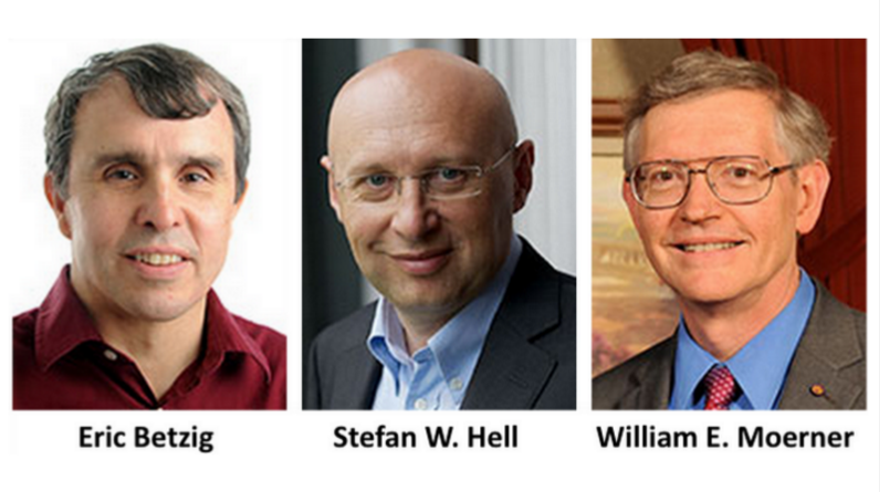
“The Nobel Prize in Chemistry 2014 was awarded jointly to Eric Betzig, Stefan W. Hell and William E. Moerner “for the development of super-resolved fluorescence microscopy”.
Source: Nobel Assembly at Karolinska Institutet
The Nobel Prize in Chemistry 2014 was awarded jointly to Eric Betzig, Stefan W. Hell and William E. Moerner”. Credits left to right: Photo- Matt Stanley/HHMI, (c) Bernd Schuller, Max Planck-Institut, Photo: K. Lowder via Wikimedia Commons, CC-BY SA 3.0
Eric Betzig website google search wikipedia entry
Stefan W. Hell website google search wikipedia entry
William E. Moerner website google search wikipedia entry
For a long time optical microscopy was held back by a presumed limitation: that it would never obtain a better resolution than half the wavelength of light. Helped by fluorescent molecules the Nobel Laureates in Chemistry 2014 ingeniously circumvented this limitation. Their ground-breaking work has brought optical microscopy into the nanodimension.
In what has become known as nanoscopy, ...
OnAir Post: 2014 Chemistry Nobel Prize
Associate Professor in Radiology, Harvard Medical School Associate Biophysicist, Massachusetts General Hospital Director, MGH NMR Core, Martinos Center
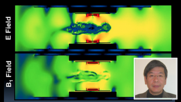
Technique development for high field imaging of the Brain. Development of 7 Tesla scanner and coils for imaging human brain function, highly parallel phased array coil development for 3T and 7T, Parallel transmit methods for B1+ mitigation in the head at 7T, and highly accelerated echo volume imaging.
Webpage: martinos.org/user/5615 Harvard Catalyst Profile: https://connects.catalyst.harvard.edu/Profiles/display/Person/42452 Neuroscience@Harvard Brain Initiative Grant
Email: wald@nmr.mgh.harvard.edu Phone: 617-724-9706 Address: Building 75, Room 2.109 13th Street Charlestown, MA 02129 USA
PhD Physics, U.C. Berkeley, 1992
Technique development for high field imaging of the Brain. Development of 7 Tesla scanner and coils for imaging human brain function, highly parallel phased array coil development for 3T and 7T, Parallel transmit methods for B1+ mitigation in the head at 7T, and highly accelerated echo volume imaging.
OnAir Post: Lawrence Wald, Phd – Harvard Med
Professor, Departments of Radiology and Biomedical Engineering, University of Minnesota Faculty, Center for Magnetic Resonance Research
Chen’s research focuses on development of magnetic resonance imaging (MRI)/spectroscopy (MRS) methodologies and technologies for noninvasively studying cellular metabolism, bioenergetics, function and dysfunction of the brain and other organs. He has been a principal investigator for a large number of NIH RO1 grants, served as grant reviewer for many funding organizations and editorial boards for imaging journals.
Webpage: cmrr.umn.edu/facultystaff/wei.shtml Institute for Translational Neuroscience Brain Initiative Grant
Email: wei@cmrr.umn.edu Phone: 612-625-8814 Address: 1-211E CMRR University of Minnesota 2021 Sixth Street SE Minneapolis, MN 55455
Dr. Chen received his B.S. degree in physical chemistry at Fudan University in Shanghai, China. In 1985, he joined Professor Ackerman’s lab as a graduate student at Washington University in St. Louis and received his Ph.D. in 1990. He spent three years as a postdoctoral fellow and research associate in Professor Shulman’s lab at Yale University Medical School. In 1994, he joined the Center for Magnetic Resonance Research (CMRR) at the University of Minnesota and became a full professor in 2002.S
Dr Chen’s research focuses on development of magnetic resonance imaging (MRI)/spectroscopy (MRS) methodologies and technologies for noninvasively studying cellular metabolism, bioenergetics, function and dysfunction of the brain and other organs. ...
OnAir Post: Wei Chen, PhD – Minnesota
