Inscopix is a discovery-phase neuroscience company in Palo Alto, CA, that develops integrated solutions for understanding the brain in action. Inscopix serves its clients in over a hundred academic and neuropharmaceutical research laboratories through its flagship brain imaging product, nVista, data analytics suite, Mosaic, and training workshops.
We have assembled here a number of videos about Inscopix: its products, people, and researchers.
Research
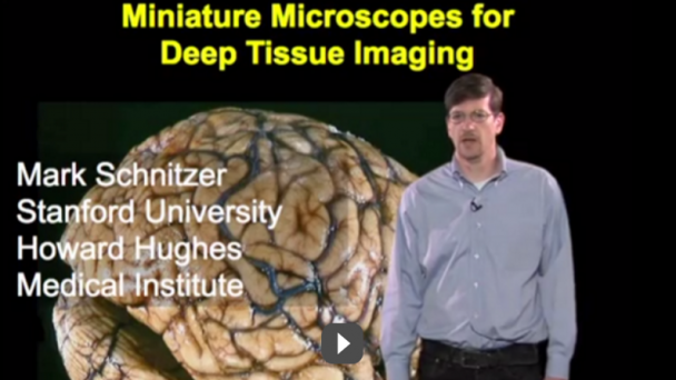
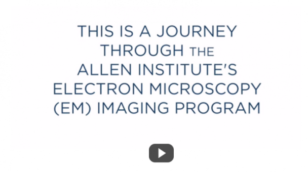
Miniature Microscopes for Deep Tissue Imaging
Published on Nov 11, 2013 iBioEducation
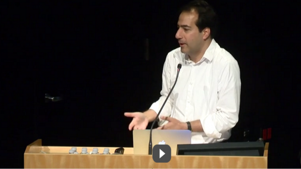
This lecture describes recent work on developing small microscopes for deep tissue imaging that can surgically implementing into living and awake animals. Exciting applications are described for imaging the activity and long term shape changes of single neurons in the brain.
https://www.youtube.com/watch?v=C1HO3ot0K00Video can’t be loaded because JavaScript is disabled: Microscopy: Miniature Microscopes for Deep Tissue Imaging (Mark Schnitzer) (https://www.youtube.com/watch?v=C1HO3ot0K00)Neocortex (Somatosensory)
Published on Jun 3, 2015 by Inscopix
OnAir Post: Inscopix and Miniature Microscopes