GE Brain Health Initiative

The Initiative will build on and coordinate multiple efforts within the company, including corporate venture capital, open innovation, R&D, and health care lines of business. This includes previously announced efforts at GE, such as the work that the company has done to convene the traumatic brain injury community and support for “Brain Trust” meetings of thought leaders.

OnAir Post: GE Brain Health Initiative

Neuroscape Lab & Glass Brain

The Neuroscape Lab is using newly emerging technology with the primary goal of driving rapid translation of neuroscience to real-world solutions. The Glass Brain visualization is one of the lab's projects.

It is being developed as a core research facility at the UCSF Neuroscience Imaging Center (NIC) under the direction of Dr. Adam Gazzaley.

OnAir Post: Neuroscape Lab & Glass Brain

IOM Workshop on Non-Invasive Neuromodulation

Based on advances in biotechnology and neuroscience, non-invasive neuromodulation devices are poised to gain clinical importance in the coming years and to be of increasing interest to patients, clinicians, health systems, payers, and industry –

Given the growing interest in non-invasive neuromodulation technologies, the Institute of Medicine’s Forum on Neuroscience and Nervous System Disorders convened a workshop in March 2015, inviting a range of stakeholders to explore the opportunities, challenges, and ethical questions surrounding the development, regulation, and reimbursement of these devices.

Figure from Dayan et al., Nature Neuroscience, 2013, showing typical noninvasive brain stimulation (NIBS) setups. (Left) Standard figure-eight TMS coil placed over dorsolateral prefrontal cortex. (Right) Bipolar tDCS electrode configuration with one electrode over dorsolateral prefrontal cortex and the other over the contralateral supraorbital region.

Report on the Workshop

Non-Invasive Neuromodulation of the Central Nervous System

To download a free PDF of Non-Invasive Neuromodulation of the Central Nervous System: Opportunities and Challenges: Workshop Summary,  log in with your existing MyNAP account, create a free MyNAP account, or download as a guest.

Walter Koroshetz summary

Noninvasive Brain Stimulation: Applications and Implications

Messages from the NINDS Director

In March the Institute of Medicine held a ...

OnAir Post: IOM Workshop on Non-Invasive Neuromodulation

Vascular Interfaces for Brain Imaging

PI: Robert Desimone Massachusetts Institute of Technology Title: “Vascular Interfaces for Brain Imaging and Stimulation” BRAIN category: Next Generation Human Imaging (RFA MH-14-217)

Dr. Desimone’s project will access the brain through its network of blood vessels to less invasively image, stimulate and monitor electrical and molecular activity than existing methods.

NIH Webpages

Sketch showing constitution of blood vessels inside the brain. Credit: Armin Kübelbeck

Project Description

Functional MRI (fMRI), EEG, and other completely noninvasive modalities for large-scale imaging of human brain activity have pioneeringly revealed many human brain functions, but cannot reach the single-neuron, single-spike level of neural code analysis possible in animals obtained using electrodes. This is partly due to the indirect methods of observation employed (e.g., blood flow for fMRI) and due to blurring of signals over distance by the skull (e.g., for EEG). In contrast, invasive approaches such as trans-cranially implanted multi- electrode arrays can achieve single-cell, single-spike resolution, but they necessitate opening of the skull – and, for implanted arrays, damage of the brain tissue – limiting utility to a small fraction of the population, those undergoing neurosurgery for some intractable brain disorder that justifies the risk. Trans-cranially implanted arrays also degrade i performance over time ...

OnAir Post: Vascular Interfaces for Brain Imaging

Ultrasonic neuromodulation in vivo

PI: Doris Ying  Tsao California Institute of Technology Title: “Dissecting human brain circuits in vivo using ultrasonic neuromodulation” BRAIN category: Next Generation Human Imaging (RFA MH-14-217)

In rodents, monkeys and eventually humans, Dr. Tsao’s team will explore use of non-invasive, high resolution ultrasound to impact neural activity deep in the brain and modify behavior.

NIH Webpages


Project Description

A dream of neuroscience is to be able to non-invasively modulate any given region of the human brain with high spatial resolution. This would open new horizons for understanding human brain function and connectivity, and create completely new options for the non-invasive treatment of brain diseases such as intractable epilepsy, depression, and Parkinson’s disease. Current non-invasive brain stimulation methods such as transcranial magnetic stimulation (TMS) and transcranial electrical stimulation (TES) can be applied only to superficial cortical areas, with crude 1 cm-scale resolution, limits placed upon these techniques by fundamental physics. Ultrasonic neuromodulation, the use of ultrasound as an energy modality to affect the activity of the brain, could overcome these limitations and thereby transform both basic and clinical human neuroscience. In fact, the engineering challenge of non-invasively focusing ultrasound to mm-sized regions, either shallow or deep in the brain, has been solved: clinical studies have already demonstrated ...

OnAir Post: Ultrasonic neuromodulation in vivo

Enhancers define cortical interneuron types

Principal Investigator: John L. R. Rubenstein UCSF Neuroscience Title: “Identification of enhancers whose activity defines cortical interneuron types” BRAIN Category: Tools for Cells and Circuits (RFA MH-14-216)

Dr. Rubenstein and colleagues plan to identify enhancer molecules specific to particular types of interneurons – that relay neural signals – and use this information to profile distinct cell types and new ways to manipulate genes.

NIH Webpages

Cortical Circuits: Projection Neurons and Interneurons

Project Description

Molecular definitions of neural cell types largely depend on the expression of RNAs or proteins as assessed by in situ hybridization, RNA array and sequencing, and immunohistochemistry. However, recent studies are demonstrating that gene regulatory elements, such as enhancers, can have highly specific spatial and temporal activity patterns in the developing brain. Thus, enhancer activity can be used to define neural cell types, and importantly, also have other broad applications. First, they can be used as tools to drive gene expression in specific cell types, which can then be used to visualize and/or purify the cells (GFP), modify gene expression in the cells (Cre), modify electrical activity (channel rhodopsin), and visualize electrical activity in the cells (GCaMP). Secondly, knowledge about the nature and position of enhancers enables geneticists ...

OnAir Post: Enhancers define cortical interneuron types

Holographic optogenetics and olfactory coding

Principal Investigator: Dmitry Rinberg NYU Neuroscience Institute Title: “Behavioral readout of spatiotemporal codes dissected by holographic optogenetics” BRAIN Category: Understanding Neural Circuits (RFA NS-14-009)

Dr. Rinberg’s team aims to understand how the brain turns odors into nerve signals by activating and recording neurons in the olfactory bulbs of mice as they detect a variety of odors.

NIH Webpages

Scientists are now able to determine which receptors respond to certain odors in awake, freely behaving animals. This image is for illustrative purposes only and shows a coronal section through the main olfactory bulb of an adult male mouse. Credit Matt Valley.

Project Description

Two of the most fundamental questions of sensory neuroscience are: 1) how is stimulus information represented by the activity of neurons at different levels of information processing? And 2) what features of this activity are read by the higher brain areas to guide behavior? The first question has been the subject of a large body of work across different sensory modalities. To answer the second question, one needs to establish a causal link between neuronal activity and behavior. In many systems, fine spatiotemporal patterns of activity underlie the neural representation of information. In these systems, deciphering the salient ...

OnAir Post: Holographic optogenetics and olfactory coding

Optogenetic toolkit for control of cells

PI: Gregory Hannon,  Hannon Lab Institution: Cold Spring Harbor Laboratory Title: “An optogenetic toolkit for the interrogation and control of single cells.” BRAIN Category: Tools for Cells and Circuits (RFA MH-14-216)

Dr. Hannon’s group will develop optogenetic techniques that use pulses of light to control genes and isolate proteins in specific cell types in the brain for molecular studies.

Project Description

Our understanding of brain function at the cellular and circuit level is critically dependent on the ability to interrogate and alter neural cells withhigh specificity. The use of light, either through single-photon or multi- photon excitation, is the onl method that provides sufficient resolution to probe the brain at the cellular and subcellular levels. While light-activated molecules, like optogenetic proteins or photocaged compounds, have allowed many key insights in neuroscience, their use is still limited to those processes that can be affected by membrane channels. We propose to develop a toolkit allowing the interrogation, regulation, and modification of genetic information in brain cells using light. We wll build on a technology we have recently developed, “LaserTag”, based on the light-dependent interaction between protein tags (i.e. SNAP-tag or HALO-tag) and caged chemical ligands. Such interaction can be either use to recover molecules through affinity purification or to force the ...

OnAir Post: Optogenetic toolkit for control of cells

Activity measurement at single cell

Principal Investigator: Craig Forest Georgia Institute of Technology Title: “In-vivo circuit activity measurement at single cell, sub-threshold resolution” BRAIN Category: Tools for Cells and Circuits (RFA MH-14-216)

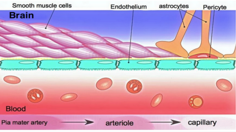
Dr. Forest’s team will use a newly developed robot guided technique to measure precise changes in electrical activity from individual neurons that are connected over long distances across the brain, to understand how these connections change when our brains go into different states, such as sleeping and waking.

NIH Webpages


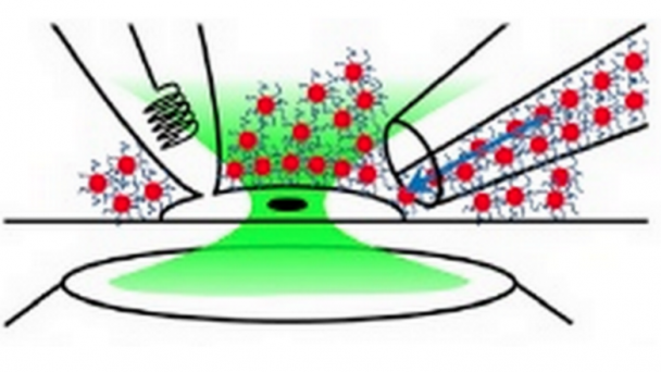
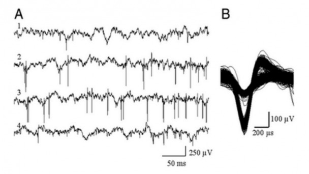
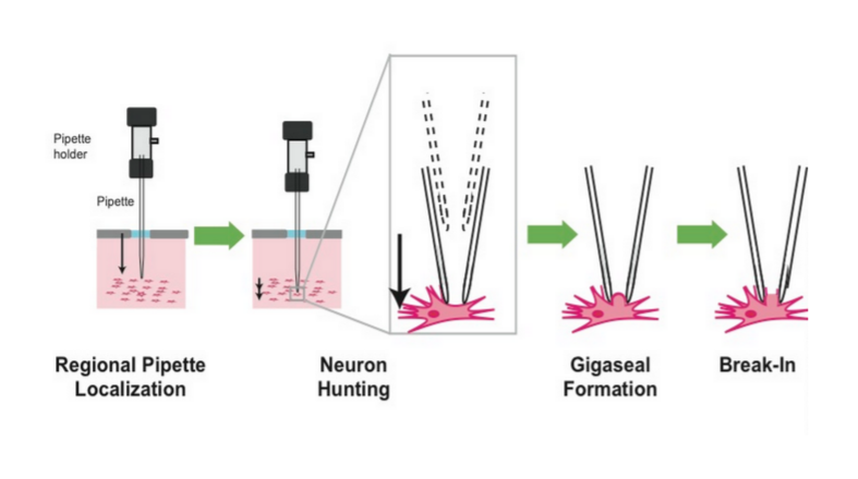
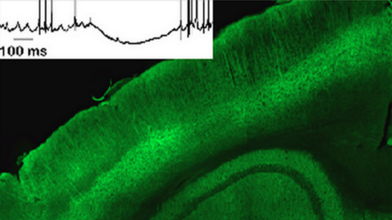
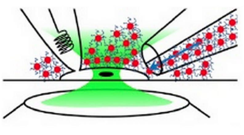
Whole-cell patch clamp electrophysiology of neurons, although a gold standard technique for high-fidelity analysis of the biophysical mechanisms of neural computation and pathology, requires great skill to perform. We have developed a simple robot that automatically performs patch clamping in vivo, algorithmically detecting cells by analyzing the temporal sequence of electrode impedance changes. We demonstrate good yield, throughput, and quality of recording in mouse cortex and hippocampus..

Project Description

Neurons communicate information through fluctuations in the electrical potentials across their cellular membranes. Whole-cell patch clamping, the gold standard technique for measuring these fluctuations, is something of an art form, requiring great skill to perform on only a few cells per day. Thus, it has been primarily limited to in vitro experiments, a few in ...

OnAir Post: Activity measurement at single cell

Artifical neuron mimics human cells

Scientists at Sweden’s Karolinska Institutet have built what they claim is a fully functional neuron by using organic bioelectronics. This artificial neuron mimics the function of a human nerve cell and communicates in the same way as neurons do.

Such a device could eventually be miniaturized and implantable, says lead investigator Agneta Richter-Dahlfors, Karolinska Institutet professor of cellular microbiology. The research objective: improve treatments for neurological disorders, which are currently limited to traditional electrical stimulation.

Chemical-to-electrical-to-chemical signal transmission. A conventional neuron (upper panel) senses chemical signals (orange circles), which trigger an electrical pulse of membrane depolarization (action potential) along the axon, causing chemical release at the axon terminals (blue circles). This process can be mimicked (lower panel) by a chemical biosensor (for glutamate or acetylcholine) connected to an axon-mimicking organic electronic ion pump that transmits electrons/ions and generates chemicals — forming an organic electronic biomimetic neuron. (credit: Daniel T. Simon et al./Biosensors and Bioelectronics

Press Release

Artifical neuron mimicks function of human cells

June 24, 2015

Scientists at Sweden’s Karolinska Institutet have managed to build a fully functional neuron by using organic bioelectronics. This artificial neuron contain no ‘living’ parts, but is capable of mimicking the ...

OnAir Post: Artifical neuron mimics human cells

Dreadd2.0: A Chemogenetic Toolkit

Principal Investigator: Bryan L Roth UNC Neuroscience Title: ” Dreadd2.0: An Enhanced Chemogenetic Toolkit” BRAIN Category: Tools for Cells and Circuits (RFA MH-14-216)

Dr. Roth and colleagues will build second generation technology that uses artificial neurotransmitters and receptors to manipulate brain activity simultaneously across select cells and pathways to understand their functions and potentially treat brain disorders.

NIH Webpages

Roth’s Lab has pioneered the use of directed molecular evolution to create GPCRs which are suitable for remotely controlling cellular signaling. Using a variety of mouse genetic approaches (e.g. Cre-mediated recombination. they are able to control neuronal firing and non-neuronal signaling in real-time in awake, freely moving animals.Ongoing projects are to use this technology to deconstruct the neuronal requirements for simple and complex behaviors, particularly as they relate to schizophrenia and drug abuse

Project Description

The Brain Research through Advancing Innovative Neurotechnologies (BRAIN) Initiative has the ambitious goal of elucidating how neuronal ensembles interactively encode higher brain processes. To accomplish this goal, new and improved methods for both recording and manipulating neuronal activity will be needed. In this application, we focus on technologies for manipulating neuronal activity. The major significance of this application is that we will provide an enhanced chemogenetic toolbox that ...

OnAir Post: Dreadd2.0: A Chemogenetic Toolkit

Ultrasound tech for degenerative diseases

https://www.youtube.com/watch?v=0ruQ7zU4yE0Video can’t be loaded because JavaScript is disabled: Ultrasound technology could treat degenerative brain diseases – Science Nation (https://www.youtube.com/watch?v=0ruQ7zU4yE0)

“Ultrasound technology could treat degenerative brain diseases”

Elisa Konofagou, a bioengineer at Columbia University, believes ultrasound technology could become be a vital component in treating and perhaps curing degenerative brain diseases such as Cancer, Alzheimer’s and Parkinson’s disease. One big problem associated with treating these diseases today is associated with the blood/brain barrier…a chemical shield of sorts that protects the brain against chemicals in the blood. Konofagou believes ultrasound waves could be one key to turning the blood/brain barrier on and off.

NSF BRAIN Initiative Science Nation – April 2, 2014

Description

Elisa Konofagou, a bioengineer at Columbia University, believes ultrasound technology could become be a vital component in treating and perhaps curing degenerative brain diseases such as Cancer, Alzheimer’s and Parkinson’s disease. One big problem associated with treating these diseases today is associated with the blood/brain barrier…a chemical shield of sorts that protects ...

OnAir Post: Ultrasound tech for degenerative diseases

Calcium sensors for molecular fMRI

PI: Alan Jasanoff Massachusetts Institute of Technology Title: “Calcium sensors for molecular fMRI” BRAIN category: Large-Scale Recording-Modulation – New Technologies (RFA NS-14-007)

Dr. Jasanoff’s team will synthesize calcium-sensing contrast agents that will allow functional magnetic resonance imaging (fMRI) scans to reveal activity of individual brain cells.

NIH Webpages

Project Description

The development of minimally invasive direct readouts of neural activity is one of the greatest challenges facing neuroscience today. Our recent work has shown that it is possible to perform high resolution functional magnetic resonance imaging (fMRI) of molecular-level phenomena using MRI contrast agents sensitive to hallmarks of neurotransmitter release. An even more valuable contribution would be the creation of calcium sensors suitable for molecular fMRI of intracellular neural signaling processes. Functional imaging performed with these sensors would combine the noninvasiveness and whole-brain coverage of MRI with the molecular specificity and broad applicability of established optical calcium neuroimaging techniques. Calcium-dependent fMRI will be a breakthrough technique for analysis of neural circuits in animals, with potential longer term applications in humans. The technique could achieve cellular resolution in conjunction with ultrahigh field MRI scanners and cell labeling techniques. A major hurdle in realizing this advance is the creation of effective calcium-dependent MRI contrast agents, however. This proposal describes ...

OnAir Post: Calcium sensors for molecular fMRI

Brain Waves: Recording from 12 neurons at once

https://www.youtube.com/watch?v=ILgONEL1Jz0Video can’t be loaded because JavaScript is disabled: Brain Waves: Recording from 12 neurons at once (https://www.youtube.com/watch?v=ILgONEL1Jz0)

The art and science of measuring the electrical activity of many individual neurons at the same time

Published on March 24, 2015  by Allen Institute for Brain Science

OnAir Post: Brain Waves: Recording from 12 neurons at once

Lloyd Watts: 2013 Allen Symposium


https://www.youtube.com/watch?v=U5-YeSZwFREVideo can’t be loaded because JavaScript is disabled: Lloyd Watts: 2013 Annual Symposium (https://www.youtube.com/watch?v=U5-YeSZwFRE)

Commercializing auditory neuroscience

Dr. Donald “Lloyd” Watts has worked as an engineer at Microtel Pacific Research, Synaptics and Arithmos. In 1997, he joined Paul Allen’s Interval Research Corporation and continued his research in reverse-engineering the human auditory pathway. In 2000, he founded Audience, Inc., to commercialize his research, with investment from Paul Allen, Carver Mead and Allan Crawford. He served as Chairman and Chief Executive Officer from 2000-2005, leading the development of the company’s core technologies. In 2005, he transitioned to the role of Audience’s Chief Technology Officer. In 2011, he became Chief Scientist. In 2013, he retired from Audience.


Published on October 3, 2013 by Allen Institute for Brain Science

OnAir Post: Lloyd Watts: 2013 Allen Symposium

Turning off Parkinson’s and depression

https://www.youtube.com/watch?v=9wiHfHOPbyEVideo can’t be loaded because JavaScript is disabled: Parkinson’s, depression and the switch that might turn them off – Andres Lozano (https://www.youtube.com/watch?v=9wiHfHOPbyE)

Deep brain stimulation is becoming very precise. This technique allows surgeons to place electrodes in almost any area of the brain, and turn them up or down — like a radio dial or thermostat — to correct dysfunction. Andres Lozano offers a dramatic look at emerging techniques, in which a woman with Parkinson’s instantly stops shaking and brain areas eroded by Alzheimer’s are brought back to life.

Filmed January 2013 at TEDs Caltech 2013 Uploaded to YouTube on June 12,, 2013 by TED 

OnAir Post: Turning off Parkinson’s and depression

Early Access to Neuromodulation and Recording

TIME: 12:00:00 PM DATE: June 3 and 4, 2015

PLACE: Neuroscience Center Building (NSC)

Live NIH Videocast (archived after seminar)

OnAir Post: Early Access to Neuromodulation and Recording

Taking genetics out of optogenetics

Light can be used to activate normal, non-genetically modified neurons through the use of targeted gold nanoparticles—a new technique that could hold promise for treating diseases such as macular degeneration.

“Many optogenetic experimental designs can now be applied to completely normal tissues or animals, greatly extending the scope of these research tools and possibly allowing for new therapies involving neuronal photostimulation.”

Neuron 2015.02.033

Funtionalized heated gold nanoparticles are not washed away, allowing them to serve a neural stimulators (credit: Joa˜ o L. Carvalho-de-Souza/Neuron)

Press Release

From University of Chicago News 3/12/15 by Kevin Jiang

New technique uses light to take genetics out of optogenetics

Gold nanoparticles enable stimulation of non-genetically modified neurons

Light can be used to activate normal, non-genetically modified neurons through the use of targeted gold nanoparticles—a new technique that could hold promise for treating diseases such as macular degeneration, scientists from the University of Chicago and the University of Illinois at Chicago report March 12 in the journal Neuron. This technique represents a significant technological advance with potential advantages over current optogenetic methods.

“This is effectively optogenetics without genetics,” said study senior author Francisco Bezanilla, the Lillian Eichelberger Cannon Professor of Biochemistry and Molecular Biology at ...

OnAir Post: Taking genetics out of optogenetics

Bioelectricity can Control Brain Development

Bioelectrical signals among cells control and instruct embryonic brain development and manipulating these signals can repair genetic defects and induce development of healthy brain tissue in locations where it would not ordinarily grow.

These bioelectric signals are implemented by changes in the voltage difference across cell membranes – called the cellular resting potential — and the patterns of differential voltages across anatomical regions.

Journal of Neuroscience 3/11/15

Left: normal tadpole brain. Center: injections with a suppressor of neural induction (Notch) caused a significantly elevated incidence of malformed brain in tadpole embryos, including near complete loss of forebrain/olfactory bulbs, and malformed midbrain and eyes. The embryos also exhibited loss of the normal voltage pattern. Right: Restoring hyperpolarization (normal voltage pattern) restored normal brain morphology, with well-formed forebrain/olfactory bulb, midbrain, and hindbrain. (credit: Vaibhav P. Pai et al./ The Journal of Neuroscience)

Press Release

Tufts University Now 3/11/15

Bioelectricity Plays Key Role in Brain Development & Repair

More than on/off switch, electric signals tell cells where & how to grow

MEDFORD/SOMERVILLE, Mass.—  Research reported today by Tufts University biologists shows for the first time that bioelectrical signals among cells control and instruct embryonic brain development and manipulating these ...

OnAir Post: Bioelectricity can Control Brain Development

3D Holography for Optogenetic Manipulation

Principal Investigator: Serge Picaud Pierre and Marie Curie University Title: “Three Dimensional Holography for Parallel Multi-target Optogenetic Circuit Manipulation” BRAIN Category: Large-Scale Recording-Modulation – Optimization (RFA NS-14-008)

Dr. Picaud’s team will continue its development of holographic imaging to use lasers to induce the natural electrical activity of neurons and test theories of how circuits produce behaviors in a range of animal models.

NIH Webpages

Zebrafish motoneurons visible thanks to a green fluorescent molecule, the GFP (Green Fluorescent Protein)© Inserm/Leclerc, Philippe

Project Description

Understanding communication between neurons, who is talking to whom, and what language they are speaking, is essential for discovering how brain circuits underlie brain function and dysfunction. Over the past decades, Neuroscience has made exponential progress toward recording and imaging communication between neurons. In addition, geneticists have recently developed the capability to manipulate neurons with light through the expression of light-activated microbial proteins called “opsins.” Now, neuroscientists can drive neural circuits in order to determine how they give rise to sensation, perception, and cognitive function. In order to take full advantage of “optogenetic” tools, we are developing holographic methods to deliver patterned light into brain tissue, to enable simultaneous activation of multiple neurons, independently controlling the strength and ...

OnAir Post: 3D Holography for Optogenetic Manipulation

Optical control of synaptic transmission

Principal Investigator: Richard  Kramer UC Berkeley Helen Wills Neuroscience Institute Title: ” Optical control of synaptic transmission for in vivo analysis of brain circuits and behavior” BRAIN Category: Large-Scale Recording-Modulation – Optimization (RFA NS-14-008)

Dr. Kramer’s team will develop light-triggered chemical compounds that selectively activate or inhibit neurotransmitter receptors on neurons, to precisely control the signals sent between brain cells in behaving animals.

NIH Webpages

Targeting Specific Channels and Receptors for Photocontrol – We have developed a strategy for photocontrol of particular ion channels and receptors with “photoswitchable tethered ligands” (PTLs).

Project Description

Optogenetics has revolutionized neuroscience by making it possible to use heterologously expressed light-gated ion channels and pumps to stimulate or inhibit action potential firing of genetically selected neurons in order to define ther roles in brain circuits and behavior. Since the flow of information through neural circuits depends on synaptic transmission between cells, an important next technological step is to bring optogenetic control to the neurotransmitter receptors of the synapse. The Optogenetic Pharmacology that we propose makes this possible. In this approach genetically-engineered neurotransmitter receptor channels and G protein coupled receptors (GCPRs) from synapse are derivatized with synthetic Photoswitched Tethered Ligands (PTLs) and thereby made controllable by light. ...

OnAir Post: Optical control of synaptic transmission

Electrophysiological Recording and Control

Principal Investigator: Baldwin Goodell Graymatter Research Title: “Large-Scale Electrophysiological Recording and Optogenetic Control System” BRAIN Category: Large-Scale Recording-Modulation – Optimization (RFA NS-14-008)

Dr. Goodell and his colleagues aim to develop optrodes, which are implantable columns of lights and wires for simultaneous electrical recording of neurons and delivery of light flashes to multiple brain areas.

NIH Webpages

Example data collected from 4 electrodes during a single recording session. A: short epoch of raw data sampled from 4 electrodes in area 7a of the posterior parietal cortex of an alert monkey. The signals are broadband (1 Hz to 10 kHz). Extracellular action potentials are visible as negative going spikes in the signals. B: plot of 1,000 superimposed waveforms extracted from the high-pass filtered signal on channel 3. The high-amplitude waveforms reveal a single unit that is well separated from the lower amplitude multiunit activity.

Project Description

In order to gain a greater understanding of the neural mechanisms that mediate human cognitive function new approaches and technologies are needed to dramatically expand the ability to record and manipulate the activity of large numbers of neurons throughout widespread areas of the primate brain. Over the past 5-10 years, our groups have made two major ...

OnAir Post: Electrophysiological Recording and Control

Modular nanophotonic probes

Principal Investigator: Michael Roukes Caltech Neuroscience Title: “Modular nanophotonic probes for dense neural recording at single-cell resolution” BRAIN Category: Large-Scale Recording-Modulation – New Technologies (RFA NS-14-007)

Dr. Roukes and his team propose to build ultra-dense, light-emitting and -sensing probes for optogenetics, which could simultaneously record the electrical activity of thousands of neurons in any given region of the brain.

NIH Webpages

Nanoprobes, Wireless, and Synthetic Biology Technologies for the BAM Project(Left) Silicon nanoprobe arrays (after Du et al., 2009b ). (A) Flip-chip assembly scheme for connecting the silicon devices with printed circuit boards. (B) SEM micrograph of the rear section of a 50-μm-thick shaft array showing the multilayer stacked structure. Adjacent layers have a spacing of 100 μm, which is set by the thickness of the flexible cable. (C) Side view of the 50-μm-thick shaft array showing that the shafts are stress balanced and are able to retain approximately constant relative spacing.(Right) Synthetic biology approaches. (D) A voltage sensitive calcium channel influences the error rate of an engineered DNA polymerase. X marks sites of mismatch between “T” in the template strand (lower) and “G” new copy strand. Note scale of the various devices and cells.

Project Description

Our understanding of ...

OnAir Post: Modular nanophotonic probes

Optogenetic mapping of synaptic activity

Principal Investigator: John Yu-Luen Lin Neuroscience at UCSD Title: “Optogenetic mapping of synaptic activity and control of intracellular signaling” BRAIN Category: Large-Scale Recording-Modulation – New Technologies (RFA NS-14-007)

Dr. Lin’s team will create molecules that, when they are triggered by a pulse of light, allow scientists to test for communication between neurons in specific circuits of the brain.

NIH Webpages

Project Description

This proposal aims to develop new molecular techniques to map activities of neurons, manipulate the strength of communication between neurons and disrupt intracellular signaling. These ‘optogenetic’ approaches will be used to further our understandings of brain function on behavior and have important implications in our understandings of neurological conditions and neurodegenerative diseases. The first goal is to develop a technique where the researchers can use optical approach to identify synaptic connections that were active during the performance of a behavior task. This reporter system can be turned on with light, which defines the window of activity reporting, and fluorescence signal can be detected if there is significant activity between two defined cell groups. Many existing approaches can only be used to map excitatory connections, whereas the proposed approach can be used to identify activities between synapses utilizing any neurotransmitters. The approach will utilize ...

OnAir Post: Optogenetic mapping of synaptic activity

Optoelectrodes for Local Circuit Analysis

Principal Investigator: Euisik  Yoon University of Michigan Neuroscience  Title: ” Modular High-Density Optoelectrodes for Local Circuit Analysis” BRAIN Category: Large-Scale Recording-Modulation – New Technologies (RFA NS-14-007)

In this project, Dr. Yoon’s team will make devices for optogenetics, a technique that enables scientists to turn neurons on and off with flashes of light, more precise and diverse by integrating multiple light sources in such a way as to enable the control of specific neuronal circuits.

NIH Webpages

Analog Front-End Module With Moderate Inversion and Power-Scalable Sampling Operation for 3-D Neural MicrosystemsWe report an analog front-end prototype designed in 0.25 CMOS process for hybrid integration into 3-D neural recording microsystems. For scaling towards massive parallel neural recording, the prototype has investigated some critical circuit challenges in power, area, interface, and modularity.

Project Description

A number of scientific questions, especially in local circuit analysis, require manipulating neurons in vivo at multiple sites independently at high spatial and temporal resolutions by perturbing a controlled number and simultaneously recorded neurons. Optogenetic stimulation is cell-type specific which has proven to be the most powerful means of circuit control. Several laboratories have developed solutions to deliver optical stimulation to deep brain structures whilst simultaneously recording neurons. However, stimulation ...

OnAir Post: Optoelectrodes for Local Circuit Analysis

Genetically encoded reporters

Principal Investigator: Kit S. Lam UC Davis Center for Neuroscience Title: “Genetically encoded reporters of integrated neural activity for functional mapping of neural circuitry” BRAIN Category: Large-Scale Recording-Modulation – New Technologies (RFA NS-14-007)

Dr. Lam’s team plans to develop fluorescent sensors that will mark ion channels, molecules that help control information flow in the brain, and enable scientists to observe the neurons that are activated during a specific behavior, such as running.

NIH Webpages

Graphic from Shultz Chemical Cell Biology Group at Heidelberg

Project Description

One of the major challenges in neuroscience is to link the structure to the function of neural circuits. To achieve this goal, we need to understand the connectivity between defined neuronal populations and the contribution of these neurons to physiological processes, behavioral responses and disease states. Recent advances in imaging techniques allow us to visualize the brain structure with cellular resolution. Application of the current generation of genetically encoded optical tools, such as sensors and controllers, is facilitating measurement and manipulation of neuron activity from molecular-defined cell populations in awake, behaving animals. However, probing the dynamics of neural circuitry underlying behavior, specifically for dissecting functional-defined circuitry beyond molecular-defined circuitry, not only depends on the improvement ...

OnAir Post: Genetically encoded reporters

Time-Reversal Optical Focusing

Principal Investigator: Changhuei Yang Caltech Neuroscience Title: Time-Reversal Optical Focusing for Noninvasive Optogenetics BRAIN Category:  Large-Scale Recording-Modulation – New Technologies (RFA NS-14-007)

Dr. Yang’s team plans to develop a light and sound system that will noninvasively shine lasers on individual cells deep within the brain and activate light-sensitive molecules to precisely guide neuronal firing.

NIH Webpages

Optofluidic microscopy (OFM) is a new compact and lensless microscopic imaging technique invented in our lab. It abandons the conventional microscope design, which requires expensive lenses and large space to magnify images, and instead utilizes microfluidic flow to deliver specimens across array(s) of micrometer-size apertures defined on a metal-coated CMOS sensor to generate direct projection images. The size of our OFM prototype device is as small as a US quarter, and yet can render images comparable in quality to those of a microscope with 20X objective.

Project Description

Our bodies appear optically opaque because biological tissue scatters light strongly. Although advances such as multiphoton excitation have enabled deeper access for optical imaging by gating out scattered light, these strategies are still fundamentally limited to superficial depths (~ 1 mm). Yang’s group at Caltech has pioneered time-reversal symmetry of optical scattering as a direct strategy ...

OnAir Post: Time-Reversal Optical Focusing

Genetic Sparse Labeling Mammalian Neuron

Principal Investigator: X. William Yang UCLA Neuroscience Title: “Novel Genetic Strategy for Sparse Labeling and Manipulation of Mammalian Neurons” BRAIN Category: Tools for Cells and Circuits (RFA MH-14-216)

Dr. Yang’s team will develop a new way to genetically target specific neurons, incorporating streamlined imaging and mapping methods that will enable the detection of sparse populations of cells that often elude existing methods.


NIH Webpages

Project Description

Cajal revolutionized the study of the brain through the use of the Golgi stain to label cells sparsely and stochastically in a fashion that revealed a neuron’s morphology in its entirety. Although genetic tools for sparse and stochastic labeling and manipulation of single neurons in Drosophila have been used extensively over the past 15 years, they have only recently become available for mammalian systems, but the latter tools are limited to only a few systems for which cell-type specific reagents (e.g. enhancers) are available or otherwise involve cumbersome manipulations. Thus, there is an important need in the field to develop robust reagents for analysis of neurons at the level of single cells. Indeed, analysis of neurons at the single identified cellular level provides critical information on the control of neuronal morphology, connectivity, physiology and plasticity. This application is in response to BRAIN ...

OnAir Post: Genetic Sparse Labeling Mammalian Neuron

Skip to toolbar